Nursing practice questions with comprehensive rationales
NurseDive Free Nursing Practice Question
I have edited the text according to your instructions. Here is the edited text and the answer for the question:
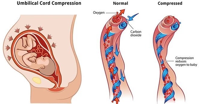
The patient is having fetal heart rate decelerations. An amnioinfusion has been ordered for the patient to alleviate the decelerations. The nurse understands that the type of decelerations that will be alleviated by amnioinfusion is:
Select one:
A. Late decelerations.
Reason: Late decelerations. This is an incorrect answer that refers to a different type of fetal heart rate patern that indicates uteroplacental insufficiency, which can reduce blood flow and oxygen delivery to the fetus. Late decelerations are characterized by gradual decreases in fetal heart rate that begin after the peak of uterine contractions and return to baseline after the end of contractions. Amnioinfusion is not effective for late decelerations, as it does not address the underlying cause of uteroplacental insufficiency, which may be due to maternal hypertension, diabetes, preeclampsia, or placental abruption.
B. Moderate decelerations.
Reason: Moderate decelerations. This is an incorrect answer that refers to a non-existent type of fetal heart rate patern, as there is no such term as moderate decelerations. The term moderate refers to the category of fetal heart rate variability, which is a measure of the fluctuations in fetal heart rate around the baseline. Moderate variability indicates normal fetal oxygenation and well-being, while absent or minimal variability indicates fetal hypoxia or distress.
C. Variable decelerations.
Reason: Variable decelerations. This is because variable decelerations are a type of fetal heart rate patern that indicates cord compression, which can reduce blood flow and oxygen delivery to the fetus. Variable decelerations are characterized by abrupt decreases in fetal heart rate that vary in onset, depth, and duration, and do not have a consistent relationship with uterine contractions. Amnioinfusion is a procedure that involves infusing saline or lactated Ringer's solution into the amniotic cavity through a transcervical catheter, which can relieve cord compression by increasing the volume of amniotic fluid and cushioning the cord. Amnioinfusion can improve fetal oxygenation and reduce variable decelerations.
D. Early decelerations.
Reason: Early decelerations. This is an incorrect answer that refers to a different type of fetal heart rate patern that indicates head compression, which can stimulate the vagus nerve and slow down the fetal heart rate. Early decelerations are characterized by gradual decreases in fetal heart rate that begin with the onset of uterine contractions and return to baseline with the end of contractions. Early decelerations are benign and do not require intervention, as they reflect normal fetal head descent and progress of labor. Amnioinfusion is not indicated for early decelerations, as it does not affect head compression or vagal stimulation.
This question is an excerpt from Nurse Dive's nursing test bank - ATI Maternity Proctored Exam 3. Take the full exam now
Full Explanation
Choice A Reason: Late decelerations. This is an incorrect answer that refers to a different type of fetal heart rate patern that indicates uteroplacental insufficiency, which can reduce blood flow and oxygen delivery to the fetus. Late decelerations are characterized by gradual decreases in fetal heart rate that begin after the peak of uterine contractions and return to baseline after the end of contractions. Amnioinfusion is not effective for late decelerations, as it does not address the underlying cause of uteroplacental insufficiency, which may be due to maternal hypertension, diabetes, preeclampsia, or placental abruption.
Choice B Reason: Moderate decelerations. This is an incorrect answer that refers to a non-existent type of fetal heart rate patern, as there is no such term as moderate decelerations. The term moderate refers to the category of fetal heart rate variability, which is a measure of the fluctuations in fetal heart rate around the baseline. Moderate variability indicates normal fetal oxygenation and well-being, while absent or minimal variability indicates fetal hypoxia or distress.
Choice C Reason: Variable decelerations. This is because variable decelerations are a type of fetal heart rate patern that indicates cord compression, which can reduce blood flow and oxygen delivery to the fetus. Variable decelerations are characterized by abrupt decreases in fetal heart rate that vary in onset, depth, and duration, and do not have a consistent relationship with uterine contractions. Amnioinfusion is a procedure that involves infusing saline or lactated Ringer's solution into the amniotic cavity through a transcervical catheter, which can relieve cord compression by increasing the volume of amniotic fluid and cushioning the cord. Amnioinfusion can improve fetal oxygenation and reduce variable decelerations.
Choice D Reason: Early decelerations. This is an incorrect answer that refers to a different type of fetal heart rate patern that indicates head compression, which can stimulate the vagus nerve and slow down the fetal heart rate. Early decelerations are characterized by gradual decreases in fetal heart rate that begin with the onset of uterine contractions and return to baseline with the end of contractions. Early decelerations are benign and do not require intervention, as they reflect normal fetal head descent and progress of labor. Amnioinfusion is not indicated for early decelerations, as it does not affect head compression or vagal stimulation.
Similar Questions
An nurse is counseling a new mother about the immunologic properties of breast milk. The nurse emphasizes that breast milk is the main source of which specific immunoglobulin?
Select one:
A. IgG.
Reason: IgG. This is an incorrect answer that refers to a different type of antibody that is not abundant in breast milk. IgG is a type of antibody that provides systemic immunity against various antigens. IgG is found in low concentrations in breast milk, as it does not cross the mammary epithelium easily. IgG can protect the infant from some infections, but it is mainly transferred from the mother to the fetus through the placenta during pregnancy.
B. IgE.
Reason: IgE. This is an incorrect answer that refers to a different type of antibody that is not relevant to breast milk. IgE is a type of antibody that mediates allergic reactions and parasitic infections. IgE is found in very low concentrations in breast milk, as it does not have a significant role in mucosal immunity. IgE can trigger mast cells and basophils to release histamine and other inflammatory mediators, which can cause symptoms such as itching, swelling, or bronchoconstriction.
C. IgA.
Reason: IgA. This is because IgA is a type of antibody that protects mucosal surfaces from pathogens and toxins. IgA is found in high concentrations in breast milk, especially in colostrum (the first milk produced after delivery). IgA can bind to bacteria and viruses in the infant's gastrointestinal tract and prevent them from ataching to the intestinal wall or crossing into the bloodstream. IgA can also enhance the infant's immune system by stimulating lymphoid tissue development and modulating inflammatory responses.
D. IgM.
Reason: IgM. This is an incorrect answer that refers to a different type of antibody that is not abundant in breast milk. IgM is a type of antibody that activates complement system and agglutinates antigens. IgM is found in low concentrations in breast milk, as it does not cross the mammary epithelium easily due to its large size. IgM can protect the infant from some infections, but it is mainly produced by the infant's own immune system in response to exposure to antigens.
Full Explanation
Choice A Reason: IgG. This is an incorrect answer that refers to a different type of antibody that is not abundant in breast milk. IgG is a type of antibody that provides systemic immunity against various antigens. IgG is found in low concentrations in breast milk, as it does not cross the mammary epithelium easily. IgG can protect the infant from some infections, but it is mainly transferred from the mother to the fetus through the placenta during pregnancy.
Choice B Reason: IgE. This is an incorrect answer that refers to a different type of antibody that is not relevant to breast milk. IgE is a type of antibody that mediates allergic reactions and parasitic infections. IgE is found in very low concentrations in breast milk, as it does not have a significant role in mucosal immunity. IgE can trigger mast cells and basophils to release histamine and other inflammatory mediators, which can cause symptoms such as itching, swelling, or bronchoconstriction.
Choice C Reason: IgA. This is because IgA is a type of antibody that protects mucosal surfaces from pathogens and toxins. IgA is found in high concentrations in breast milk, especially in colostrum (the first milk produced after delivery). IgA can bind to bacteria and viruses in the infant's gastrointestinal tract and prevent them from ataching to the intestinal wall or crossing into the bloodstream. IgA can also enhance the infant's immune system by stimulating lymphoid tissue development and modulating inflammatory responses.
Choice D Reason: IgM. This is an incorrect answer that refers to a different type of antibody that is not abundant in breast milk. IgM is a type of antibody that activates complement system and agglutinates antigens. IgM is found in low concentrations in breast milk, as it does not cross the mammary epithelium easily due to its large size. IgM can protect the infant from some infections, but it is mainly produced by the infant's own immune system in response to exposure to antigens.
Assessment of a pregnant woman reveals a pigmented vertical line between the umbilicus and the pubis. The nurse documents this finding as:
Select one:
A. Vascular spider veins.
Reason: Vascular spider veins. This is an incorrect answer that refers to a different skin change that occurs during pregnancy, which affects the blood vessels, not the pigment. Vascular spider veins are small red or purple clusters of blood vessels that appear on the skin, especially on the face, neck, chest, or legs. Vascular spider veins are caused by increased blood volume and hormonal changes, which dilate and rupture the capillaries. Vascular spider veins are harmless and usually disappear after delivery.
B. Linea nigra.
Reason: Linea nigra. This is because linea nigra is a term that refers to a darkened vertical line that appears on the abdomen during pregnancy, which runs from the umbilicus to the pubis. Linea nigra is caused by increased production of melanin, which is a pigment that gives color to the skin and hair. Linea nigra is more common and noticeable in women with darker skin tones, and it usually fades after delivery.
C. Melasma.
Reason: Melasma. This is an incorrect answer that refers to a different skin change that occurs during pregnancy, which affects the pigment, but not in a linear patern. Melasma is a term that refers to patches of brown or gray-brown discoloration that appear on the face, especially on the forehead, cheeks, nose, or upper lip. Melasma is also caused by increased production of melanin, but it is influenced by sun exposure and genetic factors. Melasma is also known as chloasma or the mask of pregnancy, and it may persist after delivery.
D. Striae gravidarum.
Reason: Striae gravidarum. This is an incorrect answer that refers to a different skin change that occurs during pregnancy, which affects the connective tissue, not the pigment. Striae gravidarum are stretch marks that appear on the skin, especially on the abdomen, breasts, hips, or thighs. Striae gravidarum are caused by rapid growth and stretching of the skin, which damage the collagen and elastin fibers. Striae gravidarum are initially red or purple, but they fade to white or silver after delivery.
Full Explanation
Choice A Reason: Vascular spider veins. This is an incorrect answer that refers to a different skin change that occurs during pregnancy, which affects the blood vessels, not the pigment. Vascular spider veins are small red or purple clusters of blood vessels that appear on the skin, especially on the face, neck, chest, or legs. Vascular spider veins are caused by increased blood volume and hormonal changes, which dilate and rupture the capillaries. Vascular spider veins are harmless and usually disappear after delivery.
Choice B Reason: Linea nigra. This is because linea nigra is a term that refers to a darkened vertical line that appears on the abdomen during pregnancy, which runs from the umbilicus to the pubis. Linea nigra is caused by increased production of melanin, which is a pigment that gives color to the skin and hair. Linea nigra is more common and noticeable in women with darker skin tones, and it usually fades after delivery.
Choice C Reason: Melasma. This is an incorrect answer that refers to a different skin change that occurs during pregnancy, which affects the pigment, but not in a linear patern. Melasma is a term that refers to patches of brown or gray-brown discoloration that appear on the face, especially on the forehead, cheeks, nose, or upper lip. Melasma is also caused by increased production of melanin, but it is influenced by sun exposure and genetic factors. Melasma is also known as chloasma or the mask of pregnancy, and it may persist after delivery.
Choice D Reason: Striae gravidarum. This is an incorrect answer that refers to a different skin change that occurs during pregnancy, which affects the connective tissue, not the pigment. Striae gravidarum are stretch marks that appear on the skin, especially on the abdomen, breasts, hips, or thighs. Striae gravidarum are caused by rapid growth and stretching of the skin, which damage the collagen and elastin fibers. Striae gravidarum are initially red or purple, but they fade to white or silver after delivery.

Before applying a cord clamp, the nurse assesses the umbilical cord for vessels. The nurse expects to find: Select one:
A. Two arteries, one vein.
Reason: Two arteries, one vein. This is because two arteries and one vein are the normal components of the umbilical cord, which is a structure that connects the fetus to the placenta and provides blood circulation between them. The umbilical cord carries oxygenated blood from the placenta to the fetus through the umbilical vein, and deoxygenated blood from the fetus to the placenta through the umbilical arteries.
B. Two veins, one artery.
Reason: Two veins, one artery. This is an incorrect answer that indicates an abnormal anatomy of the umbilical cord, which is known as single umbilical artery (SUA). SUA is a condition where there is only one umbilical artery instead of two, which can reduce blood flow and oxygen delivery to the fetus. SUA can be associated with congenital anomalies or growth restriction in some cases.
C. Two veins, two arteries.
Reason: Two veins, two arteries. This is an incorrect answer that indicates an abnormal anatomy of the umbilical cord, which is known as double umbilical vein (DUV). DUV is a condition where there are two umbilical veins instead of one, which can increase blood flow and oxygen delivery to the fetus. DUV can be associated with fetal overgrowth or polycythemia in some cases.
D. One artery, one vein.
Reason: One artery, one vein. This is an incorrect answer that indicates an abnormal anatomy of the umbilical cord, which is also known as single umbilical artery (SUA). SUA is a condition where there is only one umbilical artery instead of two, which can reduce blood flow and oxygen delivery to the fetus. SUA can be associated with congenital anomalies or growth restriction in some cases.
Full Explanation
Choice A Reason: Two arteries, one vein. This is because two arteries and one vein are the normal components of the umbilical cord, which is a structure that connects the fetus to the placenta and provides blood circulation between them. The umbilical cord carries oxygenated blood from the placenta to the fetus through the umbilical vein, and deoxygenated blood from the fetus to the placenta through the umbilical arteries.
Choice B Reason: Two veins, one artery. This is an incorrect answer that indicates an abnormal anatomy of the umbilical cord, which is known as single umbilical artery (SUA). SUA is a condition where there is only one umbilical artery instead of two, which can reduce blood flow and oxygen delivery to the fetus. SUA can be associated with congenital anomalies or growth restriction in some cases.
Choice C Reason: Two veins, two arteries. This is an incorrect answer that indicates an abnormal anatomy of the umbilical cord, which is known as double umbilical vein (DUV). DUV is a condition where there are two umbilical veins instead of one, which can increase blood flow and oxygen delivery to the fetus. DUV can be associated with fetal overgrowth or polycythemia in some cases.
Choice D Reason: One artery, one vein. This is an incorrect answer that indicates an abnormal anatomy of the umbilical cord, which is also known as single umbilical artery (SUA). SUA is a condition where there is only one umbilical artery instead of two, which can reduce blood flow and oxygen delivery to the fetus. SUA can be associated with congenital anomalies or growth restriction in some cases.