Nursing practice questions with comprehensive rationales
NurseDive Free Nursing Practice Question
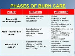
Most of the deaths from burn trauma in the emergent phase that require a referral to a burn center result from:
A. cardiac arrest related to septic shock
Cardiac arrest related to septic shock:Septic shock can occur in burn patients due to the breakdown of the skin barrier, which allows pathogens to enter the bloodstream and cause systemic infection. However, while septic shock is a serious complication of burn injuries, it is not the primary cause of death in the emergent phase. Septic shock can lead to multiple organ failure and contribute to mortality, but it is often a later complication rather than an immediate cause in the emergent phase.
B. Infection
Infection:Infections are a significant concern in burn patients, especially as the burn wound provides an ideal environment for bacterial growth. However, infections typically contribute more significantly to mortality in the later phases of burn care rather than in the emergent phase. In the emergent phase, hypovolemic shock and other immediate complications have a greater impact on mortality.
C. adrenal failure
Adrenal failure: Adrenal failure, specifically acute adrenal insufficiency or Addisonian crisis, can occur in burn patients due to the stress response and corticosteroid depletion. While adrenal insufficiency is a concern in severe burn cases, it is not the primary cause of death in the emergent phase requiring referral to a burn center.
D. hypovolemic shock and renal failure.
Hypovolemic shock and renal failure:Hypovolemic shock is a critical concern in the emergent phase of burn trauma because burns can lead to significant fluid loss and electrolyte imbalances. Hypovolemic shock results from insufficient circulating blood volume, leading to inadequate perfusion of organs and tissues, which can be life-threatening. Additionally, renal failure can develop due to hypovolemia, decreased cardiac output, and the release of inflammatory mediators, leading to acute kidney injury (AKI). Hypovolemic shock and subsequent renal failure are major contributors to mortality in the emergent phase of burn trauma, necessitating prompt referral to a burn center for specialized care.
This question is an excerpt from Nurse Dive's nursing test bank - Ati Lpn Med Surg Cohort 6 Proctored Exam. Take the full exam now
Full Explanation
A. Cardiac arrest related to septic shock:
Septic shock can occur in burn patients due to the breakdown of the skin barrier, which allows pathogens to enter the bloodstream and cause systemic infection. However, while septic shock is a serious complication of burn injuries, it is not the primary cause of death in the emergent phase. Septic shock can lead to multiple organ failure and contribute to mortality, but it is often a later complication rather than an immediate cause in the emergent phase.
B. Infection:
Infections are a significant concern in burn patients, especially as the burn wound provides an ideal environment for bacterial growth. However, infections typically contribute more significantly to mortality in the later phases of burn care rather than in the emergent phase. In the emergent phase, hypovolemic shock and other immediate complications have a greater impact on mortality.
C. Adrenal failure:
Adrenal failure, specifically acute adrenal insufficiency or Addisonian crisis, can occur in burn patients due to the stress response and corticosteroid depletion. While adrenal insufficiency is a concern in severe burn cases, it is not the primary cause of death in the emergent phase requiring referral to a burn center.
D. Hypovolemic shock and renal failure:
Hypovolemic shock is a critical concern in the emergent phase of burn trauma because burns can lead to significant fluid loss and electrolyte imbalances. Hypovolemic shock results from insufficient circulating blood volume, leading to inadequate perfusion of organs and tissues, which can be life-threatening. Additionally, renal failure can develop due to hypovolemia, decreased cardiac output, and the release of inflammatory mediators, leading to acute kidney injury (AKI). Hypovolemic shock and subsequent renal failure are major contributors to mortality in the emergent phase of burn trauma, necessitating prompt referral to a burn center for specialized care.
Similar Questions
A nurse cares for a patient who has a deep wound that is being treated with a wet to-damp (used to be dry) dressing. Which intervention would the nurse include in this patient’s plan of care?
A. Change the dressing when it is saturated.
Change the dressing when it is saturated:This intervention is the most appropriate for managing a deep wound with a wet to-damp dressing. Wet to-damp dressings are designed to maintain a moist environment conducive to wound healing. Changing the dressing when it becomes saturated with wound exudate helps prevent excessive moisture accumulation, which can lead to skin maceration and potential infection. It ensures that the wound bed remains in an optimal healing environment and reduces the risk of complications.
B. Assess the wound bed once a day.
Assess the wound bed once a day:Assessing the wound bed is an essential part of wound care, as it allows the nurse to monitor healing progress, assess for signs of infection, and evaluate the effectiveness of the chosen dressing. However, the frequency of wound bed assessment may vary depending on the specific patient's needs and the type of dressing being used. While daily assessment is generally recommended, it does not directly dictate the timing of dressing changes for wet to-damp dressings, which are primarily changed based on saturation levels.
C. Contact the provider when the dressing leaks.
Contact the provider when the dressing leaks: Contacting the provider when the dressing leaks or when there are concerns or complications is an important step in patient care. Leaking dressings can indicate issues with the dressing application, excessive wound exudate, or potential complications such as infection. It's crucial to inform the provider promptly so that appropriate interventions can be implemented, but this instruction is more reactive and does not specifically address the timing of dressing changes.
D. Change the dressing every 6 hours.
Change the dressing every 6 hours:Changing the dressing every 6 hours is not typically recommended for wet to-damp dressings unless specifically indicated based on the patient's condition and the amount of wound exudate. Frequent dressing changes can disrupt the healing process, cause unnecessary trauma to the wound bed, and increase the risk of infection. Dressing change frequency should be based on the assessment of wound exudate and the dressing's ability to maintain a moist environment.
Full Explanation
A. Change the dressing when it is saturated:
This intervention is the most appropriate for managing a deep wound with a wet to-damp dressing. Wet to-damp dressings are designed to maintain a moist environment conducive to wound healing. Changing the dressing when it becomes saturated with wound exudate helps prevent excessive moisture accumulation, which can lead to skin maceration and potential infection. It ensures that the wound bed remains in an optimal healing environment and reduces the risk of complications.
B. Assess the wound bed once a day:
Assessing the wound bed is an essential part of wound care, as it allows the nurse to monitor healing progress, assess for signs of infection, and evaluate the effectiveness of the chosen dressing. However, the frequency of wound bed assessment may vary depending on the specific patient's needs and the type of dressing being used. While daily assessment is generally recommended, it does not directly dictate the timing of dressing changes for wet to-damp dressings, which are primarily changed based on saturation levels.
C. Contact the provider when the dressing leaks:
Contacting the provider when the dressing leaks or when there are concerns or complications is an important step in patient care. Leaking dressings can indicate issues with the dressing application, excessive wound exudate, or potential complications such as infection. It's crucial to inform the provider promptly so that appropriate interventions can be implemented, but this instruction is more reactive and does not specifically address the timing of dressing changes.
D. Change the dressing every 6 hours:
Changing the dressing every 6 hours is not typically recommended for wet to-damp dressings unless specifically indicated based on the patient's condition and the amount of wound exudate. Frequent dressing changes can disrupt the healing process, cause unnecessary trauma to the wound bed, and increase the risk of infection. Dressing change frequency should be based on the assessment of wound exudate and the dressing's ability to maintain a moist environment.
A nurse evaluates the following arterial blood gas values in a patient: pH 7.48, PaO 98 mm Hg, PaCO 28 mm Hg, and HCO - 22 mEq/L (22 mmol/L). Which patient condition does the nurse correlate with these results?
A. Diarrhea and vomiting for 36 hours
Diarrhea and vomiting for 36 hours:Diarrhea and vomiting can lead to metabolic acidosis due to loss of bicarbonate and increased hydrogen ion concentration in the blood. However, the ABG values provided indicate respiratory alkalosis (high pH and low PaCO2), which is not consistent with metabolic acidosis caused by diarrhea and vomiting. Therefore, this choice does not correlate with the ABG values.
B. Chronic obstructive pulmonary disease (COPD)
Chronic obstructive pulmonary disease (COPD):COPD is a respiratory condition characterized by airflow limitation and increased airway resistance. It can lead to respiratory acidosis due to retention of carbon dioxide (PaCO2 levels would be elevated). The ABG values in the scenario show respiratory alkalosis (low PaCO2), which is the opposite of what would be expected in COPD. Therefore, this choice does not correlate with the ABG values provided.
C. Anxiety-induced hyperventilation
Anxiety-induced hyperventilation: Anxiety-induced hyperventilation is a common cause of respiratory alkalosis. During hyperventilation, there is excessive elimination of carbon dioxide (PaCO2 levels decrease), leading to an increase in pH (alkalosis). The ABG values in the scenario show a high pH (7.48) and low PaCO2 (28 mm Hg), consistent with respiratory alkalosis seen in hyperventilation due to anxiety.
D. Diabetic ketoacidosis and chronic obstructive pulmonary disease (COPD)
Diabetic ketoacidosis and chronic obstructive pulmonary disease (COPD):Diabetic ketoacidosis (DKA) is a metabolic condition characterized by hyperglycemia, ketosis, and metabolic acidosis (low pH and low bicarbonate levels). COPD, as mentioned earlier, can lead to respiratory acidosis due to retained carbon dioxide. Neither of these conditions correlates with the ABG values provided, which show respiratory alkalosis (high pH and low PaCO2). Therefore, this choice does not correlate with the ABG values.
Full Explanation
A. Diarrhea and vomiting for 36 hours:
Diarrhea and vomiting can lead to metabolic acidosis due to loss of bicarbonate and increased hydrogen ion concentration in the blood. However, the ABG values provided indicate respiratory alkalosis (high pH and low PaCO2), which is not consistent with metabolic acidosis caused by diarrhea and vomiting. Therefore, this choice does not correlate with the ABG values.
B. Chronic obstructive pulmonary disease (COPD):
COPD is a respiratory condition characterized by airflow limitation and increased airway resistance. It can lead to respiratory acidosis due to retention of carbon dioxide (PaCO2 levels would be elevated). The ABG values in the scenario show respiratory alkalosis (low PaCO2), which is the opposite of what would be expected in COPD. Therefore, this choice does not correlate with the ABG values provided.
C. Anxiety-induced hyperventilation:
Anxiety-induced hyperventilation is a common cause of respiratory alkalosis. During hyperventilation, there is excessive elimination of carbon dioxide (PaCO2 levels decrease), leading to an increase in pH (alkalosis). The ABG values in the scenario show a high pH (7.48) and low PaCO2 (28 mm Hg), consistent with respiratory alkalosis seen in hyperventilation due to anxiety.
D. Diabetic ketoacidosis and chronic obstructive pulmonary disease (COPD):
Diabetic ketoacidosis (DKA) is a metabolic condition characterized by hyperglycemia, ketosis, and metabolic acidosis (low pH and low bicarbonate levels). COPD, as mentioned earlier, can lead to respiratory acidosis due to retained carbon dioxide. Neither of these conditions correlates with the ABG values provided, which show respiratory alkalosis (high pH and low PaCO2). Therefore, this choice does not correlate with the ABG values.
A nurse assesses a patient who is admitted for treatment of fluid overload. Which manifestations does the nurse expect to find? (Select all that apply.)
A. Increased pulse rate
Increased pulse rate:This is a common manifestation of fluid overload. Excess fluid volume can lead to an increase in cardiac output, causing the heart to pump faster and resulting in an increased pulse rate.
B. Decreased blood pressure
Decreased blood pressure:Fluid overload typically leads to increased blood volume, which can initially cause an increase in blood pressure. However, as fluid overload progresses, it can lead to fluid redistribution, venous congestion, and decreased systemic vascular resistance, ultimately resulting in decreased blood pressure.
C. Skeletal muscle weakness
Skeletal muscle weakness:Skeletal muscle weakness is not a direct manifestation of fluid overload. It is more commonly associated with electrolyte imbalances, such as hypokalemia or hypomagnesemia, which can occur as a consequence of fluid shifts but are not specific to fluid overload itself.
D. Warm and pink skin
Warm and pink skin:Warm and pink skin is not typically associated with fluid overload. Instead, it is more indicative of adequate tissue perfusion and oxygenation. In fluid overload, skin changes may include edema, cool and clammy skin due to venous congestion, or signs of skin breakdown in areas of pressure.
E. Distended neck veins
Distended neck veins, specifically jugular venous distention (JVD), are commonly seen in patients with fluid overload, especially if there is right-sided heart failure or increased central venous pressure. JVD is a result of increased venous return to the heart due to fluid accumulation.
Full Explanation
A. Increased pulse rate:
This is a common manifestation of fluid overload. Excess fluid volume can lead to an increase in cardiac output, causing the heart to pump faster and resulting in an increased pulse rate.
B. Decreased blood pressure:
Fluid overload typically leads to increased blood volume, which can initially cause an increase in blood pressure. However, as fluid overload progresses, it can lead to fluid redistribution, venous congestion, and decreased systemic vascular resistance, ultimately resulting in decreased blood pressure.
C. Skeletal muscle weakness:
Skeletal muscle weakness is not a direct manifestation of fluid overload. It is more commonly associated with electrolyte imbalances, such as hypokalemia or hypomagnesemia, which can occur as a consequence of fluid shifts but are not specific to fluid overload itself.
D. Warm and pink skin:
Warm and pink skin is not typically associated with fluid overload. Instead, it is more indicative of adequate tissue perfusion and oxygenation. In fluid overload, skin changes may include edema, cool and clammy skin due to venous congestion, or signs of skin breakdown in areas of pressure.
E. Distended neck veins:
Distended neck veins, specifically jugular venous distention (JVD), are commonly seen in patients with fluid overload, especially if there is right-sided heart failure or increased central venous pressure. JVD is a result of increased venous return to the heart due to fluid accumulation.