Nursing practice questions with comprehensive rationales
NurseDive Free Nursing Practice Question
The leukocyte indicated in the image is undergoing what process?
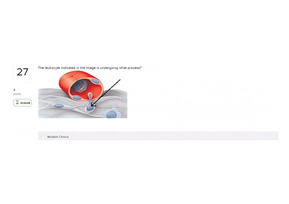
A. Diapedesis
Diapedesis (transmigration): the cell is shown squeezing through the vessel wall (leaving the bloodstream), which is diapedesis (also called extravasation).
B. Phagocytosis
Phagocytosis: phagocytosis is ingestion of particles/pathogens by a phagocyte; the image shows movement out of a vessel, not engulfment of material.
C. Chemotaxis
Chemotaxis: Incorrect (related but not the pictured action) -chemotaxis is directed movement toward chemical signals; a leukocyte may chemotax once in the tissue, but the image specifically shows the mechanical passage through the endothelium (diapedesis).
D. Margination
Margination/Rolling: margination/rolling are earlier steps along the endothelium where leukocytes slow and adhere; the image shows a cell already squeezing through the wall, which is the next step (diapedesis).
This question is an excerpt from Nurse Dive's nursing test bank - Anatomy and physiology proctored exam (Ivy college). Take the full exam now
Full Explanation
A. Diapedesis (transmigration): the cell is shown squeezing through the vessel wall (leaving the bloodstream), which is diapedesis (also called extravasation).
B. Phagocytosis: phagocytosis is ingestion of particles/pathogens by a phagocyte; the image shows movement out of a vessel, not engulfment of material.
C. Chemotaxis: Incorrect (related but not the pictured action) -chemotaxis is directed movement toward chemical signals; a leukocyte may chemotax once in the tissue, but the image specifically shows the mechanical passage through the endothelium (diapedesis).
D. Margination/Rolling: margination/rolling are earlier steps along the endothelium where leukocytes slow and adhere; the image shows a cell already squeezing through the wall, which is the next step (diapedesis).
Similar Questions
What type of antigens are found on the surface of red blood cells of a person with type AB blood?
A. Both antigens A and B
Both antigens A and B: type AB RBCs express both A and B surface antigens.
B. Neither antigens A nor antigens B
Neither antigens A nor antigens B: that describes type O, not AB.
C. Antigens A only
Antigens A only: that describes type A, not AB.
D. Antigens B only
Antigens B only: that describes type B, not AB.
Full Explanation
A. Both antigens A and B: type AB RBCs express both A and B surface antigens.
B. Neither antigens A nor antigens B: that describes type O, not AB.
C. Antigens A only: that describes type A, not AB.
D. Antigens B only: that describes type B, not AB.
Plasma electrolytes include
A. fibrinogen, globulins, and albumin
Fibrinogen, globulins, and albumin: those are plasma proteins, not electrolytes (they’re large molecules, not ions).
B. bicarbonate, magnesium, chloride, and potassium ions
Bicarbonate, magnesium, chloride, and potassium ions: these are ions dissolved in plasma and are classically considered electrolytes.
C. monocytes, basophils, and eosinophils
Monocytes, basophils, and eosinophils: those are white blood cells (formed elements), not electrolytes.
D. creatinine, urea, and uric acid
Creatinine, urea, and uric acid: those are nitrogenous waste/metabolites present in plasma, not electrolytes.
Full Explanation
A. Fibrinogen, globulins, and albumin: those are plasma proteins, not electrolytes (they’re large molecules, not ions).
B. Bicarbonate, magnesium, chloride, and potassium ions: these are ions dissolved in plasma and are classically considered electrolytes.
C. Monocytes, basophils, and eosinophils: those are white blood cells (formed elements), not electrolytes.
D. Creatinine, urea, and uric acid: those are nitrogenous waste/metabolites present in plasma, not electrolytes.
Biliverdin and bilirubin are pigments that result from the breakdown of
A. leukocytes
Leukocytes: leukocytes are white blood cells; their breakdown does not produce biliverdin/bilirubin.
B. hemoglobin
Hemoglobin: the heme portion of hemoglobin is degraded to biliverdin and then bilirubin (these are heme-breakdown pigments).
C. foreign pathogens
Foreign pathogens: pathogen breakdown is not the source of these pigments.
D. erythropoietin
Erythropoietin: erythropoietin is a hormone that stimulates RBC production; it is not broken down into biliverdin/bilirubin.
Full Explanation
A. Leukocytes: leukocytes are white blood cells; their breakdown does not produce biliverdin/bilirubin.
B. Hemoglobin: the heme portion of hemoglobin is degraded to biliverdin and then bilirubin (these are heme-breakdown pigments).
C. Foreign pathogens: pathogen breakdown is not the source of these pigments.
D. Erythropoietin: erythropoietin is a hormone that stimulates RBC production; it is not broken down into biliverdin/bilirubin.