Nursing practice questions with comprehensive rationales
NurseDive Free Nursing Practice Question
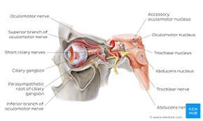
The nurse read the patient's health history and noted cranial nerve III oculomotor paralysis. Which of the following would the nurse expect?
A. The eye cannot look to the outside.
The inability of the eye to look outward, known as lateral rectus palsy, is associated with cranial nerve VI, the abducens nerve, not the oculomotor nerve. The oculomotor nerve does not control the lateral rectus muscle which governs this movement.
B. Myopia.
Myopia, or nearsightedness, is a refractive error of the eye where distant objects appear blurry while close objects can be seen clearly. It is not related to oculomotor nerve paralysis, which affects eye movements and pupil response, not the shape of the eyeball or the refractive properties of the lens.
C. Ptosis will be evident and no pupillary constriction.
Ptosis, or drooping of the upper eyelid, and an absence of pupillary constriction are classic signs of oculomotor nerve paralysis. The oculomotor nerve controls most of the eye's movements, including lifting the eyelid via the levator palpebrae superioris muscle and constricting the pupil through the circular muscles of the iris.
D. Normal eye movement.
Normal eye movement would not be expected in a patient with oculomotor nerve paralysis. This nerve controls the majority of the eye's movements, so paralysis would lead to abnormal eye movement, such as the inability to move the eye upward, downward, or inward.
This question is an excerpt from Nurse Dive's nursing test bank - Ati Fundamentals Assessment Proctored Exam Midterm. Take the full exam now
Full Explanation
Choice a reason:
The inability of the eye to look outward, known as lateral rectus palsy, is associated with cranial nerve VI, the abducens nerve, not the oculomotor nerve. The oculomotor nerve does not control the lateral rectus muscle which governs this movement.
Choice b reason:
Myopia, or nearsightedness, is a refractive error of the eye where distant objects appear blurry while close objects can be seen clearly. It is not related to oculomotor nerve paralysis, which affects eye movements and pupil response, not the shape of the eyeball or the refractive properties of the lens.
Choice c reason:
Ptosis, or drooping of the upper eyelid, and an absence of pupillary constriction are classic signs of oculomotor nerve paralysis. The oculomotor nerve controls most of the eye's movements, including lifting the eyelid via the levator palpebrae superioris muscle and constricting the pupil through the circular muscles of the iris.
Choice d reason:
Normal eye movement would not be expected in a patient with oculomotor nerve paralysis. This nerve controls the majority of the eye's movements, so paralysis would lead to abnormal eye movement, such as the inability to move the eye upward, downward, or inward.
Similar Questions
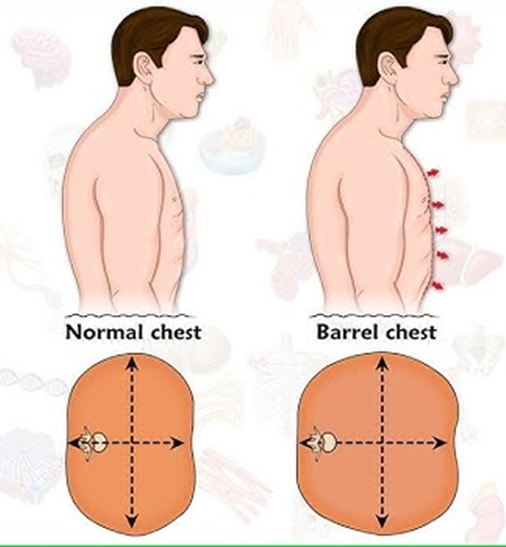
While examining a client, the nurse observes the client's chest to be barrel-shaped. The nurse would interpret this as indicating which of the following?
A. Pigeon Chest
Pigeon Chest, or pectus carinatum, is a condition where the breastbone is pushed outward, and the chest appears to protrude. It is not typically associated with a barrel-shaped chest, which is characterized by a rounded and bulging appearance.
B. Pneumonia
Pneumonia is an infection that inflames the air sacs in one or both lungs, which may fill with fluid or pus. While it can cause chest expansion, it does not lead to a barrel-shaped chest. The barrel-shaped chest is more indicative of a chronic condition rather than an acute infection like pneumonia.
C. Funnel Chest
Funnel Chest, or pectus excavatum, is a condition where the breastbone is sunken into the chest. Unlike a barrel-shaped chest, funnel chest gives the chest a depressed appearance.
D. COPD
COPD, or Chronic Obstructive Pulmonary Disease, is commonly associated with a barrel-shaped chest. This shape results from the chronic hyperinflation of the lungs due to obstructive lung disease, which causes the rib cage to remain expanded.
Full Explanation
Choice a reason:
Pigeon Chest, or pectus carinatum, is a condition where the breastbone is pushed outward, and the chest appears to protrude. It is not typically associated with a barrel-shaped chest, which is characterized by a rounded and bulging appearance.
Choice b reason:
Pneumonia is an infection that inflames the air sacs in one or both lungs, which may fill with fluid or pus. While it can cause chest expansion, it does not lead to a barrel-shaped chest. The barrel-shaped chest is more indicative of a chronic condition rather than an acute infection like pneumonia.
Choice c reason:
Funnel Chest, or pectus excavatum, is a condition where the breastbone is sunken into the chest. Unlike a barrel-shaped chest, funnel chest gives the chest a depressed appearance.
Choice d reason:
COPD, or Chronic Obstructive Pulmonary Disease, is commonly associated with a barrel-shaped chest. This shape results from the chronic hyperinflation of the lungs due to obstructive lung disease, which causes the rib cage to remain expanded.
During an assessment of the ear structures, the nurse would expect to identify which structure as part of the middle ear?
A. Tympanic Membrane
The tympanic membrane, also known as the eardrum, is a critical component of the middle ear. It is a thin membrane that separates the external ear from the middle ear and vibrates in response to sound waves. These vibrations are then transmitted to the ossicles within the middle ear, which amplify and carry the sound to the inner ear.
B. Ear lobe
The ear lobe is part of the external ear, not the middle ear. It is composed of soft skin and fatty tissue and does not play a role in hearing. The ear lobe serves primarily as a site for body decoration such as earrings.
C. Cochlea
The cochlea is a structure located in the inner ear. It is a spiral-shaped organ that contains the organ of Corti, the sensory organ of hearing. The cochlea converts the mechanical vibrations from the middle ear into nerve impulses that are sent to the brain.
D. Pinna
The pinna, or auricle, is the visible part of the external ear. It is made of cartilage and skin and functions to capture sound waves and direct them into the ear canal towards the tympanic membrane.
Full Explanation
Choice a reason:
The tympanic membrane, also known as the eardrum, is a critical component of the middle ear. It is a thin membrane that separates the external ear from the middle ear and vibrates in response to sound waves. These vibrations are then transmitted to the ossicles within the middle ear, which amplify and carry the sound to the inner ear.
Choice b reason:
The ear lobe is part of the external ear, not the middle ear. It is composed of soft skin and fatty tissue and does not play a role in hearing. The ear lobe serves primarily as a site for body decoration such as earrings.
Choice c reason:
The cochlea is a structure located in the inner ear. It is a spiral-shaped organ that contains the organ of Corti, the sensory organ of hearing. The cochlea converts the mechanical vibrations from the middle ear into nerve impulses that are sent to the brain.
Choice d reason:
The pinna, or auricle, is the visible part of the external ear. It is made of cartilage and skin and functions to capture sound waves and direct them into the ear canal towards the tympanic membrane.
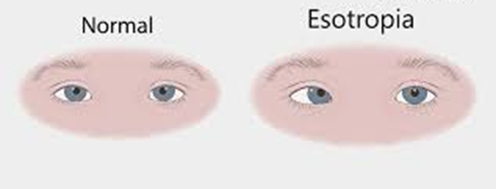
The nurse working in the eye doctor's office is completing an assessment on an elderly client. Which of the following would a nurse expect to assess in a client with esotropia?
A. Eye malalignment
Eye malalignment is a general term that refers to any form of misalignment of the eyes, which can include esotropia but is not specific to it. Esotropia is a type of strabismus where there is a specific pattern of eye malalignment.
B. Eye turning outward
Eye turning outward is known as exotropia, which is the opposite of esotropia. In exotropia, one or both eyes turn outward away from the nose, which is not characteristic of esotropia.
C. Eye oscillating
Eye oscillating refers to nystagmus, a condition where the eyes make repetitive, uncontrolled movements, often resulting in reduced vision and depth perception. While nystagmus can occur in conjunction with esotropia, it is not a defining characteristic of esotropia itself.
D. Eye turning inward
Eye turning inward is the hallmark of esotropia. In this condition, one or both eyes turn inward towards the nose. It can be constant or intermittent and may affect one eye or alternate between both eyes. Esotropia can be comitant, meaning the degree of deviation is the same in every direction of gaze, or incomitant, where the deviation varies with gaze direction.
Full Explanation
Choice a reason:
Eye malalignment is a general term that refers to any form of misalignment of the eyes, which can include esotropia but is not specific to it. Esotropia is a type of strabismus where there is a specific pattern of eye malalignment.
Choice b reason:
Eye turning outward is known as exotropia, which is the opposite of esotropia. In exotropia, one or both eyes turn outward away from the nose, which is not characteristic of esotropia.
Choice c reason:
Eye oscillating refers to nystagmus, a condition where the eyes make repetitive, uncontrolled movements, often resulting in reduced vision and depth perception. While nystagmus can occur in conjunction with esotropia, it is not a defining characteristic of esotropia itself.
Choice d reason:
Eye turning inward is the hallmark of esotropia. In this condition, one or both eyes turn inward towards the nose. It can be constant or intermittent and may affect one eye or alternate between both eyes. Esotropia can be comitant, meaning the degree of deviation is the same in every direction of gaze, or incomitant, where the deviation varies with gaze direction.