Nursing practice questions with comprehensive rationales
NurseDive Free Nursing Practice Question
What chamber of the heart is indicated in the figure?
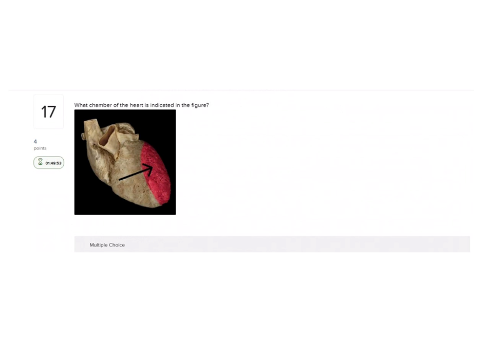
A. Right atrium
Right atrium: Receives systemic venous blood (SVC/IVC), thin-walled chamber on the heart’s right side -not the thick, muscular chamber shown if the figure indicates the left ventricle.
B. Right ventricle
Right ventricle: Pumps blood to the pulmonary trunk; its wall is thicker than an atrium but thinner than the left ventricle and sits more anteriorly -not the most muscular chamber.
C. Left atrium
Left atrium: Receives oxygenated blood from the pulmonary veins and sits posteriorly; it has thinner walls than the left ventricle.
D. Left ventricle
Left ventricle: The left ventricle has the thickest muscular wall (high systemic pressure), forms the cardiac apex, and pumps blood into the aorta
This question is an excerpt from Nurse Dive's nursing test bank - Anatomy and physiology proctored exam (Ivy college). Take the full exam now
Full Explanation
A. Right atrium: Receives systemic venous blood (SVC/IVC), thin-walled chamber on the heart’s right side -not the thick, muscular chamber shown if the figure indicates the left ventricle.
B. Right ventricle: Pumps blood to the pulmonary trunk; its wall is thicker than an atrium but thinner than the left ventricle and sits more anteriorly -not the most muscular chamber.
C. Left atrium: Receives oxygenated blood from the pulmonary veins and sits posteriorly; it has thinner walls than the left ventricle.
D. Left ventricle: The left ventricle has the thickest muscular wall (high systemic pressure), forms the cardiac apex, and pumps blood into the aorta
Similar Questions
Which cell type is an agranulocyte?
A. Monocytes
Monocytes: Agranulocyte -large cell with a kidney/bean-shaped nucleus that differentiates into macrophages in tissues.
B. Neutrophils
Neutrophils: Granulocyte -multilobed nucleus and cytoplasmic granules; primary phagocytes in acute inflammation.
C. Eosinophils
Eosinophils: Granulocyte -bilobed nucleus with large eosin-staining granules; involved in parasitic infections and allergic reactions.
D. Basophils
Basophils: Granulocyte -contains histamine/heparin-filled granules and participates in allergic/inflammatory responses.
Full Explanation
A. Monocytes: Agranulocyte -large cell with a kidney/bean-shaped nucleus that differentiates into macrophages in tissues.
B. Neutrophils: Granulocyte -multilobed nucleus and cytoplasmic granules; primary phagocytes in acute inflammation.
C. Eosinophils: Granulocyte -bilobed nucleus with large eosin-staining granules; involved in parasitic infections and allergic reactions.
D. Basophils: Granulocyte -contains histamine/heparin-filled granules and participates in allergic/inflammatory responses.
Leukocytes can undergo diapedesis. What is diapedesis?
A. The formation of a network of fibrin fibers.
The formation of a network of fibrin fibers: This describes fibrin clot formation/coagulation, not diapedesis.
B. The release of the contents of cytoplasmic granules.
The release of the contents of cytoplasmic granules: This is degranulation (e.g., in mast cells, neutrophils), not diapedesis.
C. The ability to rapidly divide, increasing the number of a particular type of leukocyte.
The ability to rapidly divide, increasing the number of a particular leukocyte: This describes cell proliferation, not diapedesis.
D. The ability of cells to squeeze between cells of capillary walls.
The ability of cells to squeeze between cells of capillary walls: Diapedesis -leukocytes migrating out of the bloodstream by squeezing between endothelial cells.
Full Explanation
A. The formation of a network of fibrin fibers: This describes fibrin clot formation/coagulation, not diapedesis.
B. The release of the contents of cytoplasmic granules: This is degranulation (e.g., in mast cells, neutrophils), not diapedesis.
C. The ability to rapidly divide, increasing the number of a particular leukocyte: This describes cell proliferation, not diapedesis.
D. The ability of cells to squeeze between cells of capillary walls: Diapedesis -leukocytes migrating out of the bloodstream by squeezing between endothelial cells.
Platelets are cell fragments that are released by what type of cell?
A. Megakaryocytes
Megakaryocytes: Megakaryocytes are large bone marrow cells whose cytoplasmic fragments become platelets (thrombocytes).
B. Macrophages
Macrophages: Phagocytic cells derived from monocytes; they do not produce platelets.
C. Monoblasts
Monoblasts: Immature precursors that develop into monocytes/macrophages, not platelets.
D. Reticulocytes
Reticulocytes: Immature red blood cells (RBC precursors) released from bone marrow—not a source of platelets.
Full Explanation
A. Megakaryocytes: Megakaryocytes are large bone marrow cells whose cytoplasmic fragments become platelets (thrombocytes).
B. Macrophages: Phagocytic cells derived from monocytes; they do not produce platelets.
C. Monoblasts: Immature precursors that develop into monocytes/macrophages, not platelets.
D. Reticulocytes: Immature red blood cells (RBC precursors) released from bone marrow—not a source of platelets.