Nursing practice questions with comprehensive rationales
NurseDive Free Nursing Practice Question
What is vasodilation?
A. Compression of a vein because of muscle contraction.
Compression of a vein because of muscle contraction: That’s mechanical venous compression (skeletal muscle pump), not vasodilation.
B. An increase in the diameter of a blood vessel.
An increase in the diameter of a blood vessel: Definition of vasodilation.
C. Contraction of the ventricles which increases ventricular pressure.
Contraction of the ventricles which increases ventricular pressure: That describes ventricular systole, not vasodilation.
D. A decrease in the diameter of a blood vessel.
A decrease in the diameter of a blood vessel: That is vasoconstriction, the opposite of vasodilation.
This question is an excerpt from Nurse Dive's nursing test bank - Anatomy and physiology proctored exam (Ivy college). Take the full exam now
Full Explanation
A. Compression of a vein because of muscle contraction: That’s mechanical venous compression (skeletal muscle pump), not vasodilation.
B. An increase in the diameter of a blood vessel: Definition of vasodilation.
C. Contraction of the ventricles which increases ventricular pressure: That describes ventricular systole, not vasodilation.
D. A decrease in the diameter of a blood vessel: That is vasoconstriction, the opposite of vasodilation.
Similar Questions
In the highlighted area, what pulse could be measured?
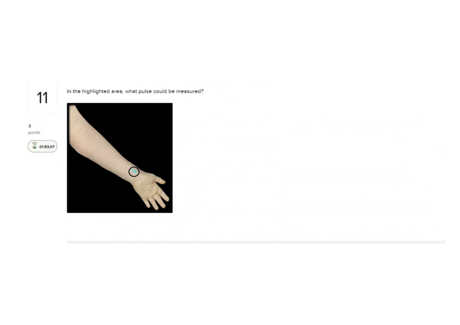
A. Radial
Radial: The radial pulse is palpated at the lateral (thumb) side of the wrist between the tendons of the flexor carpi radialis and the abductor pollicis longus -it is superficial and commonly used clinically, so this is the correct pulse for a wrist/forearm highlighted area.
B. Ulnar
Ulnar: The ulnar pulse lies on the medial (pinky) side of the wrist and is deeper and often harder to palpate than the radial; it would not be the best choice if the highlighted area is lateral at the thumb side.
C. Brachial
Brachial: The brachial pulse is felt on the medial aspect of the arm (at the antecubital fossa or just medial to the biceps tendon) and is used for infants or blood pressure -it is not located at the distal wrist.
D. Dorsalis pedis
Dorsalis pedis: The dorsalis pedis pulse is on the dorsum of the foot (between the tendons over the instep) and is unrelated to a wrist/forearm highlighting.
Full Explanation
A. Radial: The radial pulse is palpated at the lateral (thumb) side of the wrist between the tendons of the flexor carpi radialis and the abductor pollicis longus -it is superficial and commonly used clinically, so this is the correct pulse for a wrist/forearm highlighted area.
B. Ulnar: The ulnar pulse lies on the medial (pinky) side of the wrist and is deeper and often harder to palpate than the radial; it would not be the best choice if the highlighted area is lateral at the thumb side.
C. Brachial: The brachial pulse is felt on the medial aspect of the arm (at the antecubital fossa or just medial to the biceps tendon) and is used for infants or blood pressure -it is not located at the distal wrist.
D. Dorsalis pedis: The dorsalis pedis pulse is on the dorsum of the foot (between the tendons over the instep) and is unrelated to a wrist/forearm highlighting.
Which of the following would contain characteristics of cardiac muscle tissue, such as intercalated discs?
A. endocardium
Endocardium: The endocardium is the inner endothelial lining of the heart chambers (endothelium + connective tissue), not cardiac muscle, so it does not contain intercalated discs.
B. parietal pericardium
Parietal pericardium: The parietal pericardium is the outer layer of the serous/fibrous sac surrounding the heart (protective membrane), not muscle tissue, so it lacks intercalated discs.
C. epicardium
Epicardium: The epicardium (visceral layer of serous pericardium) covers the heart surface and contains connective tissue and fat; it overlies the myocardium but is not the muscle layer itself, so it does not have intercalated discs.
D. myocardium
Myocardium: The myocardium is the heart’s muscular wall composed of cardiac muscle cells that are connected by intercalated discs (for electrical and mechanical coupling).
Full Explanation
A. Endocardium: The endocardium is the inner endothelial lining of the heart chambers (endothelium + connective tissue), not cardiac muscle, so it does not contain intercalated discs.
B. Parietal pericardium: The parietal pericardium is the outer layer of the serous/fibrous sac surrounding the heart (protective membrane), not muscle tissue, so it lacks intercalated discs.
C. Epicardium: The epicardium (visceral layer of serous pericardium) covers the heart surface and contains connective tissue and fat; it overlies the myocardium but is not the muscle layer itself, so it does not have intercalated discs.
D. Myocardium: The myocardium is the heart’s muscular wall composed of cardiac muscle cells that are connected by intercalated discs (for electrical and mechanical coupling).
Select all of the accurate characteristics of veins. Check All That Apply
A. They are thicker than arteries.
They are thicker than arteries: veins generally have thinner walls (especially a thinner tunica media) than arteries because they carry blood at lower pressure.
B. Their tunica media is less developed than in arteries.
Their tunica media is less developed than in arteries: veins have a thinner tunica media (less smooth muscle and elastic tissue) compared with arteries.
C. They often have one-way valves.
They often have one-way valves: many veins (especially in the limbs) have valves to prevent backflow and assist venous return against gravity.
D. They carry blood under very high pressure.
They carry blood under very high pressure: veins return blood under low pressure; arteries carry blood under much higher pressure.
Full Explanation
A. They are thicker than arteries: veins generally have thinner walls (especially a thinner tunica media) than arteries because they carry blood at lower pressure.
B. Their tunica media is less developed than in arteries: veins have a thinner tunica media (less smooth muscle and elastic tissue) compared with arteries.
C. They often have one-way valves: many veins (especially in the limbs) have valves to prevent backflow and assist venous return against gravity.
D. They carry blood under very high pressure: veins return blood under low pressure; arteries carry blood under much higher pressure.