Nursing practice questions with comprehensive rationales
NurseDive Free Nursing Practice Question
A. A pulse of 86 beats/minute
: A pulse of 86 beats per minute is within the normal range for adults (60-100 beats per minute) and does not typically indicate a postoperative complication. While it is important to monitor vital signs, this pulse rate alone is not concerning unless accompanied by other abnormal findings.
B. Hypoactive bowel sounds in all 4 quadrants
: Hypoactive bowel sounds in all four quadrants can occur after surgery due to the effects of anesthesia and the surgical procedure itself. While it is important to monitor bowel sounds, hypoactivity is not immediately concerning unless it persists or is accompanied by other symptoms such as abdominal pain or distention. Therefore, this finding alone does not indicate an evolving complication.
C. Blood pressure of 110/70 mm Hg
: A blood pressure of 110/70 mm Hg is within the normal range for adults and does not typically indicate a postoperative complication. Blood pressure should be monitored regularly, but this reading alone is not concerning unless there are significant changes or other abnormal findings.
D. Increasing restlessness
: Increasing restlessness is a concerning sign in the immediate postoperative period. It can indicate several potential complications, including pain, hypoxia, or the onset of delirium. Restlessness may also be an early sign of shock or other serious conditions that require prompt intervention. Therefore, this symptom warrants further assessment and immediate attention to determine the underlying cause and provide appropriate treatment.
This question is an excerpt from Nurse Dive's nursing test bank - Final Med Surg Comprehensive Proctored Exam (Brooklyn University). Take the full exam now
Full Explanation
Choice A Reason:
A pulse of 86 beats per minute is within the normal range for adults (60-100 beats per minute) and does not typically indicate a postoperative complication. While it is important to monitor vital signs, this pulse rate alone is not concerning unless accompanied by other abnormal findings.
Choice B Reason:
Hypoactive bowel sounds in all four quadrants can occur after surgery due to the effects of anesthesia and the surgical procedure itself. While it is important to monitor bowel sounds, hypoactivity is not immediately concerning unless it persists or is accompanied by other symptoms such as abdominal pain or distention. Therefore, this finding alone does not indicate an evolving complication.
Choice C Reason:
A blood pressure of 110/70 mm Hg is within the normal range for adults and does not typically indicate a postoperative complication. Blood pressure should be monitored regularly, but this reading alone is not concerning unless there are significant changes or other abnormal findings.
Choice D Reason:
Increasing restlessness is a concerning sign in the immediate postoperative period. It can indicate several potential complications, including pain, hypoxia, or the onset of delirium. Restlessness may also be an early sign of shock or other serious conditions that require prompt intervention. Therefore, this symptom warrants further assessment and immediate attention to determine the underlying cause and provide appropriate treatment.
Similar Questions
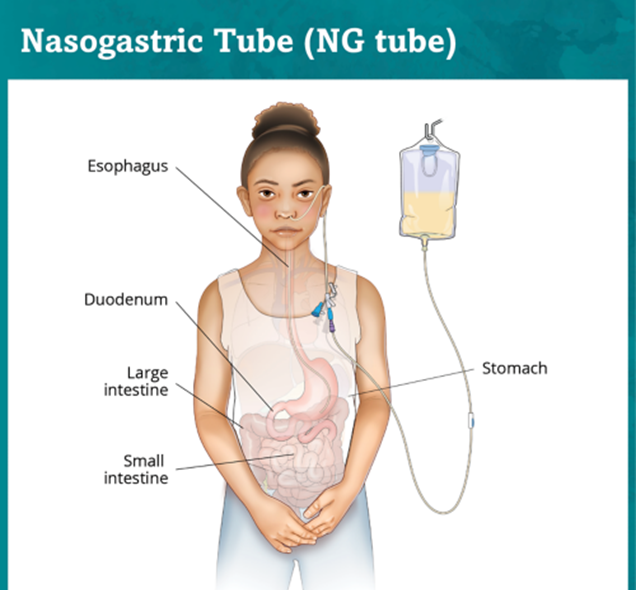
The nurse is preparing to administer medication using a client’s nasogastric tube. Which actions should the nurse take before administering the medication? Select all that apply.
A. Aspirate the stomach contents.
: Aspirating the stomach contents is essential to ensure the nasogastric tube is correctly positioned in the stomach. This step helps verify that the tube has not migrated and is safe for medication administration. If the aspirate is not obtained, further steps should be taken to confirm the tube’s placement.
B. Check the residual volume.
: Checking the residual volume is important to assess the stomach’s contents and ensure that the patient is tolerating the feedings or medications. High residual volumes may indicate delayed gastric emptying or other gastrointestinal issues. This information helps guide the timing and amount of medication administration.
C. Remove the tube and place it in the other nostril.
: Removing the tube and placing it in the other nostril is not a standard practice before administering medication. This action is unnecessary and could cause discomfort or complications for the patient. The focus should be on verifying the tube’s placement and ensuring it is functioning correctly.
D. Test the stomach contents for a pH indicating acidity.
: Testing the stomach contents for a pH indicating acidity is a reliable method to confirm the nasogastric tube’s placement. Gastric contents typically have a pH of 1 to 5, indicating the tube is in the stomach. This step helps ensure the safe administration of medications.
E. Turn off the suction to the nasogastric tube.
: Turning off the suction to the nasogastric tube is necessary before administering medications. Suction can interfere with the absorption of the medication and may cause the medication to be removed from the stomach before it has a chance to take effect. Therefore, it is important to turn off the suction temporarily during medication administration.
Full Explanation
Choice A Reason:
Aspirating the stomach contents is essential to ensure the nasogastric tube is correctly positioned in the stomach. This step helps verify that the tube has not migrated and is safe for medication administration. If the aspirate is not obtained, further steps should be taken to confirm the tube’s placement.
Choice B Reason:
Checking the residual volume is important to assess the stomach’s contents and ensure that the patient is tolerating the feedings or medications. High residual volumes may indicate delayed gastric emptying or other gastrointestinal issues. This information helps guide the timing and amount of medication administration.
Choice C Reason:
Removing the tube and placing it in the other nostril is not a standard practice before administering medication. This action is unnecessary and could cause discomfort or complications for the patient. The focus should be on verifying the tube’s placement and ensuring it is functioning correctly.
Choice D Reason:
Testing the stomach contents for a pH indicating acidity is a reliable method to confirm the nasogastric tube’s placement. Gastric contents typically have a pH of 1 to 5, indicating the tube is in the stomach. This step helps ensure the safe administration of medications.
Choice E Reason:
Turning off the suction to the nasogastric tube is necessary before administering medications. Suction can interfere with the absorption of the medication and may cause the medication to be removed from the stomach before it has a chance to take effect. Therefore, it is important to turn off the suction temporarily during medication administration.
The nurse is assessing for correct placement of a nasogastric tube. The nurse aspirates the stomach contents, checks the gastric pH, and notes a pH of 7.35. Based on this information, which action should the nurse take at this time?
A. Document that the nasogastric tube is in the correct place.
: Documenting that the nasogastric tube is in the correct place is not appropriate in this scenario. A gastric pH of 7.35 is too high for stomach contents, which typically have a pH between 1.5 and 3.5. This high pH suggests that the tube may be misplaced, possibly in the respiratory tract or another non-gastric location. Therefore, documenting the tube as correctly placed could lead to serious complications if the tube is indeed misplaced.
B. Notify the health care provider.
: Notifying the health care provider is the most appropriate action. A pH of 7.35 is indicative of a potential misplacement of the nasogastric tube. The health care provider needs to be informed immediately to take corrective actions, such as ordering an X-ray to confirm the tube’s placement or re-evaluating the tube’s position. This step is crucial to ensure patient safety and prevent complications such as aspiration pneumonia or other adverse effects.
C. Check for placement by auscultating for air injected into the tube.
: Checking for placement by auscultating for air injected into the tube is an outdated and unreliable method. This technique can sometimes give false assurance of correct placement, as the sound of air can be heard even if the tube is in the respiratory tract. Current best practices recommend using pH testing and radiographic confirmation for accurate placement verification.
D. Retest the pH using another strip.
le step, but it is not the best immediate action. If the initial pH test shows a value of 7.35, it is unlikely that retesting will yield a significantly different result. The priority should be to notify the health care provider to address the potential misplacement promptly. .
Full Explanation
Choice A Reason:
Documenting that the nasogastric tube is in the correct place is not appropriate in this scenario. A gastric pH of 7.35 is too high for stomach contents, which typically have a pH between 1.5 and 3.5. This high pH suggests that the tube may be misplaced, possibly in the respiratory tract or another non-gastric location. Therefore, documenting the tube as correctly placed could lead to serious complications if the tube is indeed misplaced.
Choice B Reason:
Notifying the health care provider is the most appropriate action. A pH of 7.35 is indicative of a potential misplacement of the nasogastric tube. The health care provider needs to be informed immediately to take corrective actions, such as ordering an X-ray to confirm the tube’s placement or re-evaluating the tube’s position. This step is crucial to ensure patient safety and prevent complications such as aspiration pneumonia or other adverse effects.
Choice C Reason:
Checking for placement by auscultating for air injected into the tube is an outdated and unreliable method. This technique can sometimes give false assurance of correct placement, as the sound of air can be heard even if the tube is in the respiratory tract. Current best practices recommend using pH testing and radiographic confirmation for accurate placement verification.
Choice D Reason:
Retesting the pH using another strip might seem like a reasonable step, but it is not the best immediate action. If the initial pH test shows a value of 7.35, it is unlikely that retesting will yield a significantly different result. The priority should be to notify the health care provider to address the potential misplacement promptly.
.
For the patient with diabetes mellitus, which microvascular complication of the blood vessel structure may occur? Select all that apply. One, some, or all responses may be correct.
A. Nephropathy
Nephropathy Diabetic nephropathy is a common microvascular complication of diabetes mellitus. It is characterized by damage to the small blood vessels in the kidneys, leading to progressive kidney disease. The condition is often identified by the presence of protein in the urine (proteinuria) and can progress to end-stage renal disease if not managed properly. The primary mechanism involves hyperglycemia-induced damage to the glomeruli, the filtering units of the kidneys. This damage results in increased permeability and eventual scarring, impairing kidney function. Effective management of blood glucose levels and blood pressure is crucial in preventing or slowing the progression of diabetic nephropathy.
B. Neuropathy
Neuropathy Diabetic neuropathy refers to nerve damage caused by chronic high blood sugar levels. It is another significant microvascular complication of diabetes. This condition can affect various types of nerves, including sensory, motor, and autonomic nerves. Symptoms may include pain, tingling, numbness, and loss of sensation, particularly in the extremities. Diabetic neuropathy can lead to severe complications such as foot ulcers and infections, which may necessitate amputation. The pathophysiology involves hyperglycemia-induced oxidative stress and inflammation, leading to nerve damage. Tight glycemic control and regular monitoring are essential in managing diabetic neuropathy.
C. Peripheral vascular disease
Peripheral Vascular Disease Peripheral vascular disease (PVD) is not classified as a microvascular complication but rather a macrovascular one. It involves the narrowing or blockage of the blood vessels outside the heart and brain, primarily affecting the arteries in the legs. PVD is associated with atherosclerosis, where plaque builds up in the arterial walls, leading to reduced blood flow. Symptoms include leg pain, cramping, and ulcers. While PVD is a significant concern for individuals with diabetes, it is not considered a microvascular complication.
D. Cerebral vascular disease
Cerebral Vascular Disease Cerebral vascular disease, which includes conditions such as stroke and transient ischemic attacks (TIAs), is also a macrovascular complication rather than a microvascular one. It involves the blood vessels supplying the brain and is primarily caused by atherosclerosis and hypertension. Diabetes increases the risk of cerebral vascular disease due to its association with other risk factors like high blood pressure and dyslipidemia. However, it is not classified as a microvascular complication.
E. Retinopathy
Retinopathy Diabetic retinopathy is a leading cause of blindness among adults with diabetes. This microvascular complication involves damage to the small blood vessels in the retina, the light-sensitive tissue at the back of the eye. There are two main types: non-proliferative and proliferative retinopathy. Non-proliferative retinopathy is characterized by microaneurysms, hemorrhages, and exudates, while proliferative retinopathy involves the growth of new, fragile blood vessels that can bleed and cause retinal detachment. The primary cause is prolonged hyperglycemia, which damages the retinal blood vessels. Regular eye examinations and good glycemic control are vital in preventing and managing diabetic retinopathy.
Full Explanation
Choice A: Nephropathy
Diabetic nephropathy is a common microvascular complication of diabetes mellitus. It is characterized by damage to the small blood vessels in the kidneys, leading to progressive kidney disease. The condition is often identified by the presence of protein in the urine (proteinuria) and can progress to end-stage renal disease if not managed properly. The primary mechanism involves hyperglycemia-induced damage to the glomeruli, the filtering units of the kidneys. This damage results in increased permeability and eventual scarring, impairing kidney function. Effective management of blood glucose levels and blood pressure is crucial in preventing or slowing the progression of diabetic nephropathy.
Choice B: Neuropathy
Diabetic neuropathy refers to nerve damage caused by chronic high blood sugar levels. It is another significant microvascular complication of diabetes. This condition can affect various types of nerves, including sensory, motor, and autonomic nerves. Symptoms may include pain, tingling, numbness, and loss of sensation, particularly in the extremities. Diabetic neuropathy can lead to severe complications such as foot ulcers and infections, which may necessitate amputation. The pathophysiology involves hyperglycemia-induced oxidative stress and inflammation, leading to nerve damage. Tight glycemic control and regular monitoring are essential in managing diabetic neuropathy.
Choice E: Retinopathy
Diabetic retinopathy is a leading cause of blindness among adults with diabetes. This microvascular complication involves damage to the small blood vessels in the retina, the light-sensitive tissue at the back of the eye. There are two main types: non-proliferative and proliferative retinopathy. Non-proliferative retinopathy is characterized by microaneurysms, hemorrhages, and exudates, while proliferative retinopathy involves the growth of new, fragile blood vessels that can bleed and cause retinal detachment. The primary cause is prolonged hyperglycemia, which damages the retinal blood vessels. Regular eye examinations and good glycemic control are vital in preventing and managing diabetic retinopathy.
Choice C: Peripheral Vascular Disease
Peripheral vascular disease (PVD) is not classified as a microvascular complication but rather a macrovascular one. It involves the narrowing or blockage of the blood vessels outside the heart and brain, primarily affecting the arteries in the legs. PVD is associated with atherosclerosis, where plaque builds up in the arterial walls, leading to reduced blood flow. Symptoms include leg pain, cramping, and ulcers. While PVD is a significant concern for individuals with diabetes, it is not considered a microvascular complication.
Choice D: Cerebral Vascular Disease
Cerebral vascular disease, which includes conditions such as stroke and transient ischemic attacks (TIAs), is also a macrovascular complication rather than a microvascular one. It involves the blood vessels supplying the brain and is primarily caused by atherosclerosis and hypertension. Diabetes increases the risk of cerebral vascular disease due to its association with other risk factors like high blood pressure and dyslipidemia. However, it is not classified as a microvascular complication.