Nursing practice questions with comprehensive rationales
NurseDive Free Nursing Practice Question
Which of the following are pancreatic secretions? (Select all that apply)
A. Pepsin
Pepsin: Pepsin is an active protease produced in the stomach (from pepsinogen) -not a pancreatic secretion.
B. Sodium bicarbonate
Sodium bicarbonate: The pancreas secretes bicarbonate-rich fluid to neutralize acidic chyme in the duodenum -pancreatic secretion .
C. Amylase
Amylase: Pancreatic amylase digests starches into sugars in the small intestine -pancreatic secretion .
D. Nuclease
Nuclease: The pancreas secretes nucleases (e.g., deoxyribonuclease, ribonuclease) to digest nucleic acids -pancreatic secretion .
E. Lipase
Lipase: Pancreatic lipase hydrolyzes triglycerides into free fatty acids and monoglycerides -pancreatic secretion .
F. Pepsinogen
Pepsinogen: Pepsinogen is secreted by chief cells in the stomach, not by the pancreas -not a pancreatic secretion.
This question is an excerpt from Nurse Dive's nursing test bank - HUMAN ANATOMY AND PHYSIOLOGY II PROCTORED EXAM (ARIZONA COLLEGE). Take the full exam now
Full Explanation
A. Pepsin: Pepsin is an active protease produced in the stomach (from pepsinogen) -not a pancreatic secretion.
B. Sodium bicarbonate: The pancreas secretes bicarbonate-rich fluid to neutralize acidic chyme in the duodenum -pancreatic secretion .
C. Amylase: Pancreatic amylase digests starches into sugars in the small intestine -pancreatic secretion .
D. Nuclease: The pancreas secretes nucleases (e.g., deoxyribonuclease, ribonuclease) to digest nucleic acids -pancreatic secretion .
E. Lipase: Pancreatic lipase hydrolyzes triglycerides into free fatty acids and monoglycerides -pancreatic secretion .
F. Pepsinogen: Pepsinogen is secreted by chief cells in the stomach, not by the pancreas -not a pancreatic secretion.
Similar Questions
Letter C indicates what part of the nephron?
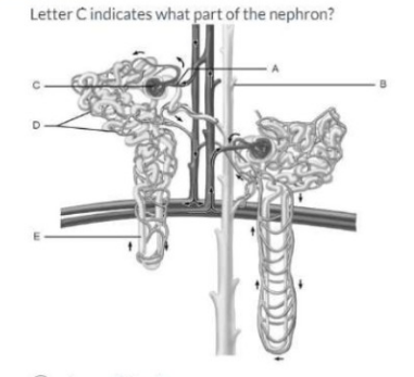
A. Loop of Henle
Loop of Henle: The Loop of Henle is the U-shaped segment of the nephron that extends into the medulla and concentrates urine.
B. Proximal Convoluted Tubule
Proximal Convoluted Tubule: The PCT is the first convoluted segment after Bowman's capsule and is responsible for bulk reabsorption of solutes and water.
C. Bowman's Capsule
Bowman's Capsule: Bowman's capsule is the double-walled cup that surrounds the glomerular capillary tuft and collects filtrate.
D. Glomerulus
Glomerulus: The glomerulus is the capillary tuft inside Bowman's capsule that performs plasma filtration.
E. Collecting Duct
The collecting duct collects filtrate from multiple nephrons and participates in final water/solute adjustments.
Full Explanation
A. Loop of Henle: The Loop of Henle is the U-shaped segment of the nephron that extends into the medulla and concentrates urine.
B. Proximal Convoluted Tubule: The PCT is the first convoluted segment after Bowman's capsule and is responsible for bulk reabsorption of solutes and water.
C. Bowman's Capsule: Bowman's capsule is the double-walled cup that surrounds the glomerular capillary tuft and collects filtrate.
D. Glomerulus: The glomerulus is the capillary tuft inside Bowman's capsule that performs plasma filtration.
E. Collecting Duct: The collecting duct collects filtrate from multiple nephrons and participates in final water/solute adjustments.
How is the small intestine adapted for nutrient absorption?
A. CCK is released from the duodenum to promote liver and pancreatic secretion.
CCK is released from the duodenum to promote liver and pancreatic secretion.: CCK (cholecystokinin) is released by duodenal/jejunal enteroendocrine cells and stimulates pancreatic enzyme secretion and gallbladder contraction -this supports digestion but describes a hormonal control mechanism rather than a structural adaptation of the small intestine itself.
B. Large surface area due to the presence of the plicae circulares, villi, and microvilli.
Large surface area due to the presence of the plicae circulares, villi, and microvilli.: The small intestine’s mucosal folds (plicae circulares), finger-like villi, and microscopic microvilli (brush border) massively increase surface area for absorption.
C. Acid secretions from the stomach are neutralized in the duodenum.
Acid secretions from the stomach are neutralized in the duodenum.: The duodenum receives bicarbonate-rich pancreatic secretions and bile which neutralize gastric acid to create an optimal pH for intestinal enzymes -this is an important functional adaptation that facilitates digestion/absorption but is secondary to the structural surface-area adaptations.
D. The small intestine has haustra and rugae both help in nutrient absorption.
The small intestine has haustra and rugae both help in nutrient absorption.: Haustra are sacculations of the large intestine (colon) and rugae are folds in the stomach -neither are features of the small intestine, so this statement is incorrect.
Full Explanation
A. CCK is released from the duodenum to promote liver and pancreatic secretion.: CCK (cholecystokinin) is released by duodenal/jejunal enteroendocrine cells and stimulates pancreatic enzyme secretion and gallbladder contraction -this supports digestion but describes a hormonal control mechanism rather than a structural adaptation of the small intestine itself.
B. Large surface area due to the presence of the plicae circulares, villi, and microvilli.: The small intestine’s mucosal folds (plicae circulares), finger-like villi, and microscopic microvilli (brush border) massively increase surface area for absorption.
C. Acid secretions from the stomach are neutralized in the duodenum.: The duodenum receives bicarbonate-rich pancreatic secretions and bile which neutralize gastric acid to create an optimal pH for intestinal enzymes -this is an important functional adaptation that facilitates digestion/absorption but is secondary to the structural surface-area adaptations.
D. The small intestine has haustra and rugae both help in nutrient absorption.: Haustra are sacculations of the large intestine (colon) and rugae are folds in the stomach -neither are features of the small intestine, so this statement is incorrect.
Name the structure labeled #3 on the following image:
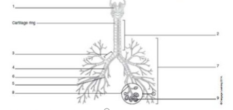
A. Primary bronchus
Primary bronchus: The primary (main) bronchus is the first large airway branch that arises at the carina from the trachea and conducts air into each lung.
B. Respiratory bronchiole
Respiratory bronchiole: Respiratory bronchioles are very small distal airways that contain alveoli and are deep within the lung parenchyma; they are much smaller than the structure shown near the main branch.
C. Trachea
Trachea: The trachea is the central airway above the bifurcation (a vertical tube with cartilage rings); a label on the long central tube would indicate the trachea, not the branching airway.
D. Secondary bronchus
Secondary bronchus: Secondary (lobar) bronchi are branches of the primary bronchus that lead to lung lobes; they are a generation distal to the main bronchus and are smaller than the primary bronchus.
Full Explanation
A. Primary bronchus: The primary (main) bronchus is the first large airway branch that arises at the carina from the trachea and conducts air into each lung.
B. Respiratory bronchiole: Respiratory bronchioles are very small distal airways that contain alveoli and are deep within the lung parenchyma; they are much smaller than the structure shown near the main branch.
C. Trachea: The trachea is the central airway above the bifurcation (a vertical tube with cartilage rings); a label on the long central tube would indicate the trachea, not the branching airway.
D. Secondary bronchus: Secondary (lobar) bronchi are branches of the primary bronchus that lead to lung lobes; they are a generation distal to the main bronchus and are smaller than the primary bronchus.