Nursing practice questions with comprehensive rationales
NurseDive Free Nursing Practice Question
Which of the following parts of an EKG occurs during ventricular depolarization?
A. QRS complex
QRS complex: The QRS represents the rapid depolarization of the right and left ventricles (electrical activity that triggers ventricular contraction).
B. T wave
T wave: The T wave represents ventricular repolarization (the ventricles recovering electrically), not depolarization.
C. P wave
P wave: The P wave represents atrial depolarization, the electrical activation of the atria.
D. PQ segment
PQ segment: The PQ (PR) segment is the brief isoelectric period while the impulse travels through the AV node/AV bundle (a conduction delay), not ventricular depolarization.
This question is an excerpt from Nurse Dive's nursing test bank - Anatomy and physiology proctored exam (Ivy college). Take the full exam now
Full Explanation
A. QRS complex: The QRS represents the rapid depolarization of the right and left ventricles (electrical activity that triggers ventricular contraction).
B. T wave: The T wave represents ventricular repolarization (the ventricles recovering electrically), not depolarization.
C. P wave: The P wave represents atrial depolarization, the electrical activation of the atria.
D. PQ segment: The PQ (PR) segment is the brief isoelectric period while the impulse travels through the AV node/AV bundle (a conduction delay), not ventricular depolarization.
Similar Questions
What chamber of the heart is indicated in the figure?
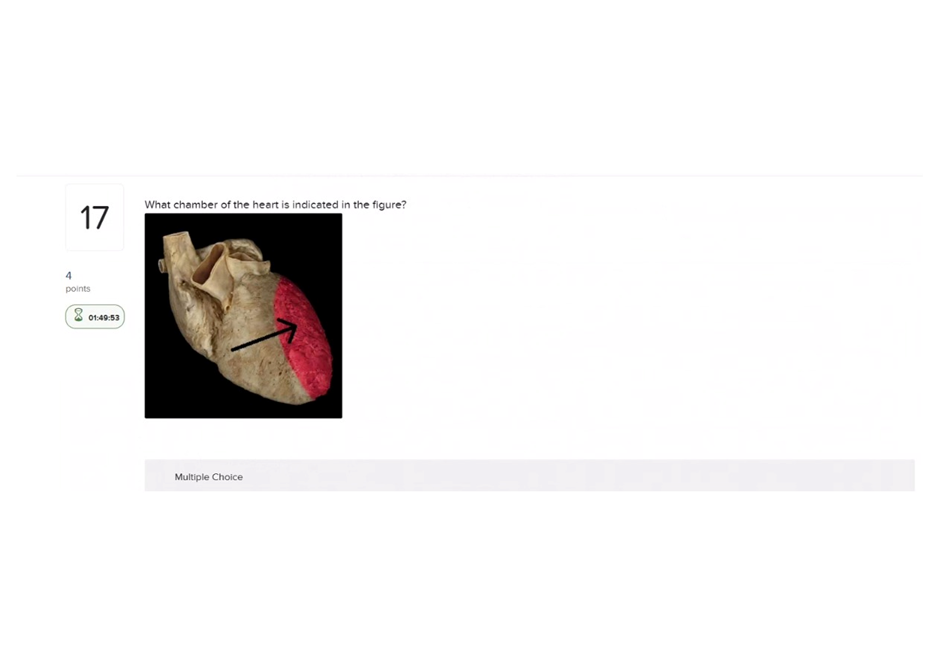
A. Right atrium
Right atrium: Receives systemic venous blood (SVC/IVC), thin-walled chamber on the heart’s right side -not the thick, muscular chamber shown if the figure indicates the left ventricle.
B. Right ventricle
Right ventricle: Pumps blood to the pulmonary trunk; its wall is thicker than an atrium but thinner than the left ventricle and sits more anteriorly -not the most muscular chamber.
C. Left atrium
Left atrium: Receives oxygenated blood from the pulmonary veins and sits posteriorly; it has thinner walls than the left ventricle.
D. Left ventricle
Left ventricle: The left ventricle has the thickest muscular wall (high systemic pressure), forms the cardiac apex, and pumps blood into the aorta
Full Explanation
A. Right atrium: Receives systemic venous blood (SVC/IVC), thin-walled chamber on the heart’s right side -not the thick, muscular chamber shown if the figure indicates the left ventricle.
B. Right ventricle: Pumps blood to the pulmonary trunk; its wall is thicker than an atrium but thinner than the left ventricle and sits more anteriorly -not the most muscular chamber.
C. Left atrium: Receives oxygenated blood from the pulmonary veins and sits posteriorly; it has thinner walls than the left ventricle.
D. Left ventricle: The left ventricle has the thickest muscular wall (high systemic pressure), forms the cardiac apex, and pumps blood into the aorta
Which cell type is an agranulocyte?
A. Monocytes
Monocytes: Agranulocyte -large cell with a kidney/bean-shaped nucleus that differentiates into macrophages in tissues.
B. Neutrophils
Neutrophils: Granulocyte -multilobed nucleus and cytoplasmic granules; primary phagocytes in acute inflammation.
C. Eosinophils
Eosinophils: Granulocyte -bilobed nucleus with large eosin-staining granules; involved in parasitic infections and allergic reactions.
D. Basophils
Basophils: Granulocyte -contains histamine/heparin-filled granules and participates in allergic/inflammatory responses.
Full Explanation
A. Monocytes: Agranulocyte -large cell with a kidney/bean-shaped nucleus that differentiates into macrophages in tissues.
B. Neutrophils: Granulocyte -multilobed nucleus and cytoplasmic granules; primary phagocytes in acute inflammation.
C. Eosinophils: Granulocyte -bilobed nucleus with large eosin-staining granules; involved in parasitic infections and allergic reactions.
D. Basophils: Granulocyte -contains histamine/heparin-filled granules and participates in allergic/inflammatory responses.
Leukocytes can undergo diapedesis. What is diapedesis?
A. The formation of a network of fibrin fibers.
The formation of a network of fibrin fibers: This describes fibrin clot formation/coagulation, not diapedesis.
B. The release of the contents of cytoplasmic granules.
The release of the contents of cytoplasmic granules: This is degranulation (e.g., in mast cells, neutrophils), not diapedesis.
C. The ability to rapidly divide, increasing the number of a particular type of leukocyte.
The ability to rapidly divide, increasing the number of a particular leukocyte: This describes cell proliferation, not diapedesis.
D. The ability of cells to squeeze between cells of capillary walls.
The ability of cells to squeeze between cells of capillary walls: Diapedesis -leukocytes migrating out of the bloodstream by squeezing between endothelial cells.
Full Explanation
A. The formation of a network of fibrin fibers: This describes fibrin clot formation/coagulation, not diapedesis.
B. The release of the contents of cytoplasmic granules: This is degranulation (e.g., in mast cells, neutrophils), not diapedesis.
C. The ability to rapidly divide, increasing the number of a particular leukocyte: This describes cell proliferation, not diapedesis.
D. The ability of cells to squeeze between cells of capillary walls: Diapedesis -leukocytes migrating out of the bloodstream by squeezing between endothelial cells.