Nursing practice questions with comprehensive rationales
NurseDive Free Nursing Practice Question
A. No change to the heparin rate
No change to the heparin rate is not appropriate in this scenario. The normal range for PTT is generally between 25 to 35 seconds. However, for a client on heparin therapy, the target PTT is typically 1.5 to 2.5 times the normal range, which would be approximately 60 to 80 seconds. Since the client’s PTT is only 25 seconds, it indicates that the blood is clotting too quickly, and the heparin dose is insufficient.
B. Decrease the heparin rate
Decreasing the heparin rate would further reduce the anticoagulant effect, which is not advisable given the current PTT of 25 seconds. Lowering the heparin rate could increase the risk of thrombus formation and worsen the deep vein thrombosis (DVT) condition.
C. Stop heparin and start warfarin
Stopping heparin and starting warfarin is not an immediate solution. Warfarin takes several days to achieve its full anticoagulant effect, and during this transition period, the client would be at risk of clot formation. Heparin provides immediate anticoagulation, which is crucial in the acute management of DVT.
D. Increase the heparin rate
Increasing the heparin rate is the correct action. The current PTT of 25 seconds is below the therapeutic range for a client on heparin therapy. Increasing the heparin rate will help achieve the desired anticoagulant effect, prolonging the PTT to the target range of 60 to 80 seconds.
This question is an excerpt from Nurse Dive's nursing test bank - Final Med Surg Comprehensive Proctored Exam (Brooklyn University). Take the full exam now
Full Explanation
Choice A reason: No change to the heparin rate is not appropriate in this scenario. The normal range for PTT is generally between 25 to 35 seconds. However, for a client on heparin therapy, the target PTT is typically 1.5 to 2.5 times the normal range, which would be approximately 60 to 80 seconds. Since the client’s PTT is only 25 seconds, it indicates that the blood is clotting too quickly, and the heparin dose is insufficient.
Choice B reason: Decreasing the heparin rate would further reduce the anticoagulant effect, which is not advisable given the current PTT of 25 seconds. Lowering the heparin rate could increase the risk of thrombus formation and worsen the deep vein thrombosis (DVT) condition.
Choice C reason: Stopping heparin and starting warfarin is not an immediate solution. Warfarin takes several days to achieve its full anticoagulant effect, and during this transition period, the client would be at risk of clot formation. Heparin provides immediate anticoagulation, which is crucial in the acute management of DVT.
Choice D reason: Increasing the heparin rate is the correct action. The current PTT of 25 seconds is below the therapeutic range for a client on heparin therapy. Increasing the heparin rate will help achieve the desired anticoagulant effect, prolonging the PTT to the target range of 60 to 80 seconds.
Similar Questions
A nurse is caring for a client who returns to the nursing unit from the recovery room after a sigmoid colon resection for adenocarcinoma. The client had an episode of intraoperative bleeding. Which finding indicates to the nurse that the client may be developing hypovolemic shock?
A. Increase in the temperature from 37.5°C (99.5°F) to 38.6°C (101.5°F)
An increase in temperature is not a primary indicator of hypovolemic shock. While fever can occur due to infection or inflammation, it is not directly related to hypovolemic shock, which is primarily characterized by a significant loss of blood or fluids leading to decreased perfusion and oxygenation of tissues.
B. Decrease in the urinary output from 50 to 30 mL per hour
A decrease in urinary output is a critical sign of hypovolemic shock. When the body loses a significant amount of blood or fluids, the kidneys receive less blood flow, leading to reduced urine production. This is a compensatory mechanism to conserve fluids and maintain blood pressure. Normal urine output is typically around 30 to 50 mL per hour, so a drop below this range is concerning.
C. Increase in the heart rate from 88 to 110/min
An increase in heart rate is a common response to hypovolemic shock as the body attempts to maintain cardiac output and blood pressure despite the loss of blood volume. Tachycardia (increased heart rate) is one of the early signs of shock, indicating that the heart is working harder to pump blood to vital organs.
D. Decrease in the respiratory rate from 20 to 16/min
A decrease in respiratory rate is not typical of hypovolemic shock. In fact, hypovolemic shock often leads to an increased respiratory rate (tachypnea) as the body tries to compensate for decreased oxygen delivery to tissues. A decrease in respiratory rate could indicate other issues but is not a hallmark of hypovolemic shock.
Full Explanation
Choice A reason: An increase in temperature is not a primary indicator of hypovolemic shock. While fever can occur due to infection or inflammation, it is not directly related to hypovolemic shock, which is primarily characterized by a significant loss of blood or fluids leading to decreased perfusion and oxygenation of tissues.
Choice B reason: A decrease in urinary output is a critical sign of hypovolemic shock. When the body loses a significant amount of blood or fluids, the kidneys receive less blood flow, leading to reduced urine production. This is a compensatory mechanism to conserve fluids and maintain blood pressure. Normal urine output is typically around 30 to 50 mL per hour, so a drop below this range is concerning.
Choice C reason: An increase in heart rate is a common response to hypovolemic shock as the body attempts to maintain cardiac output and blood pressure despite the loss of blood volume. Tachycardia (increased heart rate) is one of the early signs of shock, indicating that the heart is working harder to pump blood to vital organs.
Choice D reason: A decrease in respiratory rate is not typical of hypovolemic shock. In fact, hypovolemic shock often leads to an increased respiratory rate (tachypnea) as the body tries to compensate for decreased oxygen delivery to tissues. A decrease in respiratory rate could indicate other issues but is not a hallmark of hypovolemic shock.
A nurse is caring for a client who has Cushing’s syndrome. The nurse should recognize that which of the following are manifestations of Cushing’s syndrome? (Select all that apply)
A. Buffalo hump
A buffalo hump is a characteristic sign of Cushing’s syndrome. It refers to the accumulation of fat on the back of the neck and shoulders. This symptom occurs due to the excessive production of cortisol, which leads to abnormal fat distribution in the body.
B. Moon face
Moon face is another hallmark of Cushing’s syndrome. It describes the rounding and fullness of the face, which results from fat deposits. This symptom is also caused by prolonged exposure to high levels of cortisol.
C. Hypertension
Hypertension, or high blood pressure, is commonly associated with Cushing’s syndrome. Cortisol increases blood pressure by enhancing the sensitivity of blood vessels to catecholamines and by promoting sodium and water retention.
D. Purple striations
Purple striations, or stretch marks, are often seen in individuals with Cushing’s syndrome. These marks typically appear on the abdomen, thighs, breasts, and arms. They result from the thinning of the skin and the breakdown of collagen due to elevated cortisol levels.
E. Tremors
Tremors are not typically associated with Cushing’s syndrome. While Cushing’s syndrome can cause a variety of symptoms, tremors are more commonly linked to other conditions such as hyperthyroidism or neurological disorders.
Full Explanation
Choice A reason: A buffalo hump is a characteristic sign of Cushing’s syndrome. It refers to the accumulation of fat on the back of the neck and shoulders. This symptom occurs due to the excessive production of cortisol, which leads to abnormal fat distribution in the body.
Choice B reason: Moon face is another hallmark of Cushing’s syndrome. It describes the rounding and fullness of the face, which results from fat deposits. This symptom is also caused by prolonged exposure to high levels of cortisol.
Choice C reason: Hypertension, or high blood pressure, is commonly associated with Cushing’s syndrome. Cortisol increases blood pressure by enhancing the sensitivity of blood vessels to catecholamines and by promoting sodium and water retention.
Choice D reason: Purple striations, or stretch marks, are often seen in individuals with Cushing’s syndrome. These marks typically appear on the abdomen, thighs, breasts, and arms. They result from the thinning of the skin and the breakdown of collagen due to elevated cortisol levels.
Choice E reason: Tremors are not typically associated with Cushing’s syndrome. While Cushing’s syndrome can cause a variety of symptoms, tremors are more commonly linked to other conditions such as hyperthyroidism or neurological disorders.
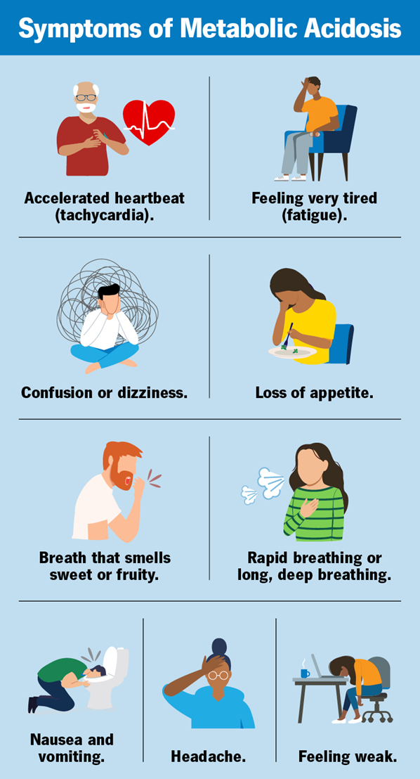
A nurse is caring for a client who has the following arterial blood gas results: HCO3 18 mEq/L, PaCO2 28 mm Hg, and pH 7.30. The nurse recognizes the client is experiencing which of the following acid-base imbalances?
A. Metabolic alkalosis
: Metabolic alkalosis is characterized by an elevated pH (greater than 7.45) and an increased bicarbonate (HCO3) level. In this case, the pH is 7.30, indicating acidosis, and the HCO3 level is 18 mEq/L, which is below the normal range (22-26 mEq/L). Therefore, metabolic alkalosis is not the correct diagnosis.
B. Respiratory alkalosis
: Respiratory alkalosis is indicated by a high pH (greater than 7.45) and a low PaCO2 (less than 35 mm Hg). Although the PaCO2 is low at 28 mm Hg, the pH is 7.30, indicating acidosis rather than alkalosis. Therefore, respiratory alkalosis is not the correct diagnosis.
C. Respiratory acidosis
: Respiratory acidosis is characterized by a low pH (less than 7.35) and an elevated PaCO2 (greater than 45 mm Hg). In this case, the pH is low, indicating acidosis, but the PaCO2 is also low at 28 mm Hg, which does not fit the criteria for respiratory acidosis. Therefore, respiratory acidosis is not the correct diagnosis.
D. Metabolic acidosis
: Metabolic acidosis is indicated by a low pH (less than 7.35) and a low bicarbonate (HCO3) level (less than 22 mEq/L). In this case, the pH is 7.30, indicating acidosis, and the HCO3 level is 18 mEq/L, which is below the normal range. The low PaCO2 of 28 mm Hg suggests a compensatory respiratory response to the metabolic acidosis. Therefore, metabolic acidosis is the correct diagnosis.
Full Explanation
Choice A Reason:
Metabolic alkalosis is characterized by an elevated pH (greater than 7.45) and an increased bicarbonate (HCO3) level. In this case, the pH is 7.30, indicating acidosis, and the HCO3 level is 18 mEq/L, which is below the normal range (22-26 mEq/L). Therefore, metabolic alkalosis is not the correct diagnosis.
Choice B Reason:
Respiratory alkalosis is indicated by a high pH (greater than 7.45) and a low PaCO2 (less than 35 mm Hg). Although the PaCO2 is low at 28 mm Hg, the pH is 7.30, indicating acidosis rather than alkalosis. Therefore, respiratory alkalosis is not the correct diagnosis.
Choice C Reason:
Respiratory acidosis is characterized by a low pH (less than 7.35) and an elevated PaCO2 (greater than 45 mm Hg). In this case, the pH is low, indicating acidosis, but the PaCO2 is also low at 28 mm Hg, which does not fit the criteria for respiratory acidosis. Therefore, respiratory acidosis is not the correct diagnosis.
Choice D Reason:
Metabolic acidosis is indicated by a low pH (less than 7.35) and a low bicarbonate (HCO3) level (less than 22 mEq/L). In this case, the pH is 7.30, indicating acidosis, and the HCO3 level is 18 mEq/L, which is below the normal range. The low PaCO2 of 28 mm Hg suggests a compensatory respiratory response to the metabolic acidosis. Therefore, metabolic acidosis is the correct diagnosis.