Nursing practice questions with comprehensive rationales
NurseDive Free Nursing Practice Question
A nurse is caring for a client with a new skin inflammation on the chest. Which of the following is an important assessment question to ask the client?
A. How many people live in your home?
How many people live in your home?This question pertains to social and environmental factors but is not directly related to assessing skin inflammation on the chest. While social factors can impact overall health, such as stress levels or exposure to infectious agents, the number of people living in the client's home is unlikely to be directly related to a new skin inflammation unless there are specific circumstances, such as sharing personal care products or close contact with others who have similar skin issues.
B. Did you have a recent exposure to Irritants?
Did you have a recent exposure to irritants?This question is highly relevant to assessing a new skin inflammation on the chest. Exposure to irritants or allergens can trigger or worsen skin conditions, such as contact dermatitis or allergic reactions. By asking about recent exposure to potential irritants like new detergents, soaps, fabrics, chemicals, or environmental factors, the nurse can gather important information to identify possible triggers for the skin inflammation.
C. Is nausea associated with your rash7
Is nausea associated with your rash? Nausea is typically not directly associated with a skin rash or inflammation unless there is a systemic condition or allergic reaction causing both symptoms. While it's important to assess for any systemic signs or symptoms that may be related to the skin condition, such as fever or malaise, specifically asking about nausea may not provide relevant information about the skin inflammation on the chest.
D. What is your body mass index?
What is your body mass index?Body mass index (BMI) is a measure of body weight relative to height and is not directly related to assessing a new skin inflammation on the chest. While obesity or changes in body weight can sometimes contribute to skin issues, such as friction-related dermatitis or hormonal changes affecting skin health, BMI alone is not a primary assessment parameter for localized skin conditions unless there are specific concerns related to weight-related skin problems.
This question is an excerpt from Nurse Dive's nursing test bank - Ati Lpn Med Surg Cohort 6 Proctored Exam. Take the full exam now
Full Explanation
-
A. How many people live in your home?
This question pertains to social and environmental factors but is not directly related to assessing skin inflammation on the chest. While social factors can impact overall health, such as stress levels or exposure to infectious agents, the number of people living in the client's home is unlikely to be directly related to a new skin inflammation unless there are specific circumstances, such as sharing personal care products or close contact with others who have similar skin issues.
B. Did you have a recent exposure to irritants?
This question is highly relevant to assessing a new skin inflammation on the chest. Exposure to irritants or allergens can trigger or worsen skin conditions, such as contact dermatitis or allergic reactions. By asking about recent exposure to potential irritants like new detergents, soaps, fabrics, chemicals, or environmental factors, the nurse can gather important information to identify possible triggers for the skin inflammation.
C. Is nausea associated with your rash?
Nausea is typically not directly associated with a skin rash or inflammation unless there is a systemic condition or allergic reaction causing both symptoms. While it's important to assess for any systemic signs or symptoms that may be related to the skin condition, such as fever or malaise, specifically asking about nausea may not provide relevant information about the skin inflammation on the chest.
D. What is your body mass index?
Body mass index (BMI) is a measure of body weight relative to height and is not directly related to assessing a new skin inflammation on the chest. While obesity or changes in body weight can sometimes contribute to skin issues, such as friction-related dermatitis or hormonal changes affecting skin health, BMI alone is not a primary assessment parameter for localized skin conditions unless there are specific concerns related to weight-related skin problems.
Similar Questions
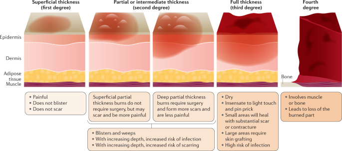
A nurse is providing education to a community group about burn prevention. Which of the following is an example of a first-degree burn?
A. Excessive scarring
Excessive scarring:Excessive scarring is not an example of a first-degree burn. It typically occurs in more severe burns that affect deeper layers of the skin, such as second-degree or third-degree burns. Second-degree burns extend into the dermis, while third-degree burns damage all layers of the skin and can lead to significant scarring. First-degree burns, on the other hand, only affect the outer layer of the skin (epidermis) and usually do not result in excessive scarring.
B. Blistering from flames
Blistering from flames:Blistering from flames is more characteristic of a second-degree burn rather than a first-degree burn. Second-degree burns involve damage to both the epidermis and part of the dermis, which can result in blister formation. These burns are often caused by direct contact with flames, hot liquids, or steam.
C. Blackened dead skin
Blackened dead skin: Blackened dead skin is indicative of a third-degree burn, which is the most severe type of burn. Third-degree burns damage all layers of the skin, including the epidermis, dermis, and sometimes underlying tissues. The skin may appear charred or blackened, and these burns often require medical intervention, such as skin grafting, due to the extent of tissue damage.
D. A sunburn
A sunburn:A sunburn is an example of a first-degree burn. It occurs due to overexposure to ultraviolet (UV) radiation from the sun, leading to redness, pain, and mild swelling of the skin. First-degree burns affect only the outer layer of the skin (epidermis) and typically heal within a few days without significant scarring or blistering. Applying soothing lotions, staying hydrated, and avoiding further sun exposure can help manage sunburns.
Full Explanation
A. Excessive scarring:
Excessive scarring is not an example of a first-degree burn. It typically occurs in more severe burns that affect deeper layers of the skin, such as second-degree or third-degree burns. Second-degree burns extend into the dermis, while third-degree burns damage all layers of the skin and can lead to significant scarring. First-degree burns, on the other hand, only affect the outer layer of the skin (epidermis) and usually do not result in excessive scarring.
B. Blistering from flames:
Blistering from flames is more characteristic of a second-degree burn rather than a first-degree burn. Second-degree burns involve damage to both the epidermis and part of the dermis, which can result in blister formation. These burns are often caused by direct contact with flames, hot liquids, or steam.
C. Blackened dead skin:
Blackened dead skin is indicative of a third-degree burn, which is the most severe type of burn. Third-degree burns damage all layers of the skin, including the epidermis, dermis, and sometimes underlying tissues. The skin may appear charred or blackened, and these burns often require medical intervention, such as skin grafting, due to the extent of tissue damage.
D. A sunburn:
A sunburn is an example of a first-degree burn. It occurs due to overexposure to ultraviolet (UV) radiation from the sun, leading to redness, pain, and mild swelling of the skin. First-degree burns affect only the outer layer of the skin (epidermis) and typically heal within a few days without significant scarring or blistering. Applying soothing lotions, staying hydrated, and avoiding further sun exposure can help manage sunburns.
The nurse is caring for a client with a chronic wound. Which of the following are wound treatments that may assist with the healing process?
A. Measure the depth and width of the wound.
Measure the depth and width of the wound. Regular assessment and documentation of the wound’s size can help track the progress of healing and effectiveness of the treatment plan.
B. Educate the client about the need for antibiotics.
Educate the client about the need for antibiotics. If an infection is present, antibiotics may be necessary. It’s important for the client to understand the purpose and proper use of these medications.
C. Consult a nutritionist for a diet plan.
Consult a nutritionist for a diet plan. Good nutrition is essential for wound healing. Certain nutrients, like protein, vitamin C, and zinc, can promote wound healing.
D. Remove any non-viable tissue.
Remove any non-viable tissue. Debridement, or the removal of dead (non-viable) tissue, can help promote the healing of the wound by reducing the risk of infection and allowing healthy tissue to grow.
Full Explanation
-
A. Measure the depth and width of the wound. Regular assessment and documentation of the wound’s size can help track the progress of healing and effectiveness of the treatment plan.
B. Educate the client about the need for antibiotics. If an infection is present, antibiotics may be necessary. It’s important for the client to understand the purpose and proper use of these medications.
C. Consult a nutritionist for a diet plan. Good nutrition is essential for wound healing. Certain nutrients, like protein, vitamin C, and zinc, can promote wound healing.
D. Remove any non-viable tissue. Debridement, or the removal of dead (non-viable) tissue, can help promote the healing of the wound by reducing the risk of infection and allowing healthy tissue to grow.
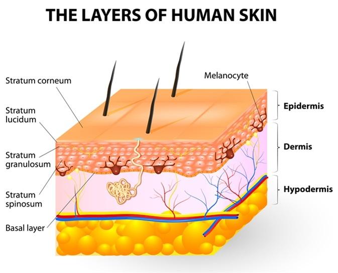
Melanocytes give rise to the pigment melanin, which is responsible for skin color. Where can the melanocytes be found?
A. Loose connective tissue
Loose connective tissue:Melanocytes are not typically found in loose connective tissue. Their primary location is within the epidermis, specifically in the basal layer, where they interact with keratinocytes to produce melanin and contribute to skin color. Loose connective tissue contains collagen and elastin fibers, as well as fibroblasts, but it does not house melanocytes.
B. Epidermis
Epidermis: This is the correct answer. Melanocytes are primarily located in the basal layer of the epidermis, which is the deepest layer of the epidermis. These cells produce melanin, a pigment that helps protect the skin from UV radiation and determines skin color. Melanocytes are interspersed among keratinocytes in the epidermis and transfer melanin to keratinocytes to provide skin pigmentation.
C. Dermis
Dermis:The dermis is the layer of skin beneath the epidermis and consists of connective tissue, blood vessels, nerves, hair follicles, and sweat glands. While the dermis plays a crucial role in supporting and nourishing the epidermis, melanocytes are not primarily located in the dermis. They are confined to the basal layer of the epidermis.
D. Superficial fascia
Superficial fascia:The superficial fascia, also known as the subcutaneous tissue or hypodermis, lies beneath the dermis and consists of adipose (fat) tissue and connective tissue. It provides insulation, energy storage, and cushioning for underlying structures. However, melanocytes are not typically found in the superficial fascia. They are restricted to the epidermis, specifically the basal layer, where they carry out their function of melanin production.
Full Explanation
A. Loose connective tissue:
Melanocytes are not typically found in loose connective tissue. Their primary location is within the epidermis, specifically in the basal layer, where they interact with keratinocytes to produce melanin and contribute to skin color. Loose connective tissue contains collagen and elastin fibers, as well as fibroblasts, but it does not house melanocytes.
B. Epidermis:
This is the correct answer. Melanocytes are primarily located in the basal layer of the epidermis, which is the deepest layer of the epidermis. These cells produce melanin, a pigment that helps protect the skin from UV radiation and determines skin color. Melanocytes are interspersed among keratinocytes in the epidermis and transfer melanin to keratinocytes to provide skin pigmentation.
C. Dermis:
The dermis is the layer of skin beneath the epidermis and consists of connective tissue, blood vessels, nerves, hair follicles, and sweat glands. While the dermis plays a crucial role in supporting and nourishing the epidermis, melanocytes are not primarily located in the dermis. They are confined to the basal layer of the epidermis.
D. Superficial fascia:
The superficial fascia, also known as the subcutaneous tissue or hypodermis, lies beneath the dermis and consists of adipose (fat) tissue and connective tissue. It provides insulation, energy storage, and cushioning for underlying structures. However, melanocytes are not typically found in the superficial fascia. They are restricted to the epidermis, specifically the basal layer, where they carry out their function of melanin production.