Nursing practice questions with comprehensive rationales
NurseDive Free Nursing Practice Question
From the left ventricle, blood is pushed into which vessel?
A. pulmonary veins
Pulmonary veins: pulmonary veins carry oxygenated blood from the lungs into the left atrium, not out of the left ventricle.
B. superior vena cava
Superior vena cava: the superior vena cava returns deoxygenated blood from the upper body to the right atrium, not from the left ventricle.
C. pulmonary trunk
Pulmonary trunk: the pulmonary trunk arises from the right ventricle and carries deoxygenated blood to the lungs.
D. aorta
Aorta: the aorta is the large artery that emerges from the left ventricle and distributes oxygenated blood to the systemic circulation.
This question is an excerpt from Nurse Dive's nursing test bank - Anatomy and physiology proctored exam (Ivy college). Take the full exam now
Full Explanation
A. Pulmonary veins: pulmonary veins carry oxygenated blood from the lungs into the left atrium, not out of the left ventricle.
B. Superior vena cava: the superior vena cava returns deoxygenated blood from the upper body to the right atrium, not from the left ventricle.
C. Pulmonary trunk: the pulmonary trunk arises from the right ventricle and carries deoxygenated blood to the lungs.
D. Aorta: the aorta is the large artery that emerges from the left ventricle and distributes oxygenated blood to the systemic circulation.
Similar Questions
What vessel drains the blood from the face and scalp?
A. External jugular vein
External jugular vein: the external jugular drains superficial structures of the head and neck (including much of the face and scalp) and empties into the subclavian vein.
B. Superior vena cava
Superior vena cava: Incorrect (in the direct sense) -the superior vena cava is the large trunk that returns venous blood to the right atrium, but it does not directly drain the face/scalp; it receives blood ultimately via the brachiocephalic veins.
C. Subclavian vein
Subclavian vein: Partially related but not the primary answer -the subclavian receives blood from the external jugular, but it primarily drains the upper limb; it is a larger trunk rather than the direct superficial drain of face/scalp.
D. Cephalic vein
Cephalic vein: the cephalic vein is a superficial vein of the lateral upper limb (arm/forearm), not a drain of the head or face.
Full Explanation
A. External jugular vein: the external jugular drains superficial structures of the head and neck (including much of the face and scalp) and empties into the subclavian vein.
B. Superior vena cava: Incorrect (in the direct sense) -the superior vena cava is the large trunk that returns venous blood to the right atrium, but it does not directly drain the face/scalp; it receives blood ultimately via the brachiocephalic veins.
C. Subclavian vein: Partially related but not the primary answer -the subclavian receives blood from the external jugular, but it primarily drains the upper limb; it is a larger trunk rather than the direct superficial drain of face/scalp.
D. Cephalic vein: the cephalic vein is a superficial vein of the lateral upper limb (arm/forearm), not a drain of the head or face.
Which of the following parts of an EKG occurs during ventricular depolarization?
A. QRS complex
QRS complex: The QRS represents the rapid depolarization of the right and left ventricles (electrical activity that triggers ventricular contraction).
B. T wave
T wave: The T wave represents ventricular repolarization (the ventricles recovering electrically), not depolarization.
C. P wave
P wave: The P wave represents atrial depolarization, the electrical activation of the atria.
D. PQ segment
PQ segment: The PQ (PR) segment is the brief isoelectric period while the impulse travels through the AV node/AV bundle (a conduction delay), not ventricular depolarization.
Full Explanation
A. QRS complex: The QRS represents the rapid depolarization of the right and left ventricles (electrical activity that triggers ventricular contraction).
B. T wave: The T wave represents ventricular repolarization (the ventricles recovering electrically), not depolarization.
C. P wave: The P wave represents atrial depolarization, the electrical activation of the atria.
D. PQ segment: The PQ (PR) segment is the brief isoelectric period while the impulse travels through the AV node/AV bundle (a conduction delay), not ventricular depolarization.
What chamber of the heart is indicated in the figure?
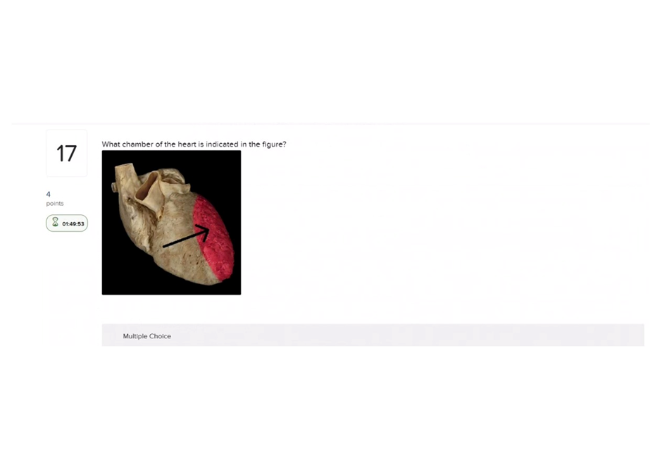
A. Right atrium
Right atrium: Receives systemic venous blood (SVC/IVC), thin-walled chamber on the heart’s right side -not the thick, muscular chamber shown if the figure indicates the left ventricle.
B. Right ventricle
Right ventricle: Pumps blood to the pulmonary trunk; its wall is thicker than an atrium but thinner than the left ventricle and sits more anteriorly -not the most muscular chamber.
C. Left atrium
Left atrium: Receives oxygenated blood from the pulmonary veins and sits posteriorly; it has thinner walls than the left ventricle.
D. Left ventricle
Left ventricle: The left ventricle has the thickest muscular wall (high systemic pressure), forms the cardiac apex, and pumps blood into the aorta
Full Explanation
A. Right atrium: Receives systemic venous blood (SVC/IVC), thin-walled chamber on the heart’s right side -not the thick, muscular chamber shown if the figure indicates the left ventricle.
B. Right ventricle: Pumps blood to the pulmonary trunk; its wall is thicker than an atrium but thinner than the left ventricle and sits more anteriorly -not the most muscular chamber.
C. Left atrium: Receives oxygenated blood from the pulmonary veins and sits posteriorly; it has thinner walls than the left ventricle.
D. Left ventricle: The left ventricle has the thickest muscular wall (high systemic pressure), forms the cardiac apex, and pumps blood into the aorta