Nursing practice questions with comprehensive rationales
NurseDive Free Nursing Practice Question
The hormone
This question is an excerpt from Nurse Dive's nursing test bank - Anatomy and physiology proctored exam (Ivy college). Take the full exam now
Full Explanation
A. Colony-stimulating factor; negative: colony-stimulating factors (CSFs) chiefly stimulate white blood cell production, not RBCs. The feedback characterization here is not applicable to erythropoiesis.
B. Erythropoietin; positive: erythropoietin (EPO) does stimulate RBC production, but the regulatory loop is negative feedback. So labeling it “positive” is wrong.
C. Erythropoietin; negative: EPO is the hormone that stimulates RBC production and it is regulated by a negative feedback mechanism based on tissue oxygenation.
D. Colony-stimulating factor; positive: CSFs affect leukocyte lines, and erythropoiesis is not regulated by a positive-CSF loop.
Similar Questions
What are the formed elements?
A. Bone marrow and the thymus
Bone marrow and the thymus: those are organs/tissues involved in blood cell production/maturation, not the formed elements themselves.
B. Blood and lymph
Blood and lymph: these are fluid compartments, not the formed cellular elements suspended in blood.
C. Blood cells and platelets
Blood cells and platelets: “formed elements” refers to erythrocytes, leukocytes, and platelets (the cellular/fragment components of blood).
D. Sodium and potassium
Sodium and potassium: those are electrolytes/ions in plasma, not formed cellular elements.
Full Explanation
A. Bone marrow and the thymus: those are organs/tissues involved in blood cell production/maturation, not the formed elements themselves.
B. Blood and lymph: these are fluid compartments, not the formed cellular elements suspended in blood.
C. Blood cells and platelets: “formed elements” refers to erythrocytes, leukocytes, and platelets (the cellular/fragment components of blood).
D. Sodium and potassium: those are electrolytes/ions in plasma, not formed cellular elements.
Iron is a component of what substance?
A. Biliverdin
Biliverdin: biliverdin is a green pigment formed during heme breakdown and does not contain iron.
B. Heme
Heme: the heme moiety of hemoglobin contains an iron atom (Fe²⁺) at its center that binds oxygen.
C. Bilirubin
Bilirubin: bilirubin is a breakdown product of biliverdin and does not contain iron.
D. Globin
Globin: globin is the protein part of hemoglobin (amino-acid chains); iron is in the heme portion, not the globin polypeptide.
Full Explanation
A. Biliverdin: biliverdin is a green pigment formed during heme breakdown and does not contain iron.
B. Heme: the heme moiety of hemoglobin contains an iron atom (Fe²⁺) at its center that binds oxygen.
C. Bilirubin: bilirubin is a breakdown product of biliverdin and does not contain iron.
D. Globin: globin is the protein part of hemoglobin (amino-acid chains); iron is in the heme portion, not the globin polypeptide.
The leukocyte indicated in the image is undergoing what process?
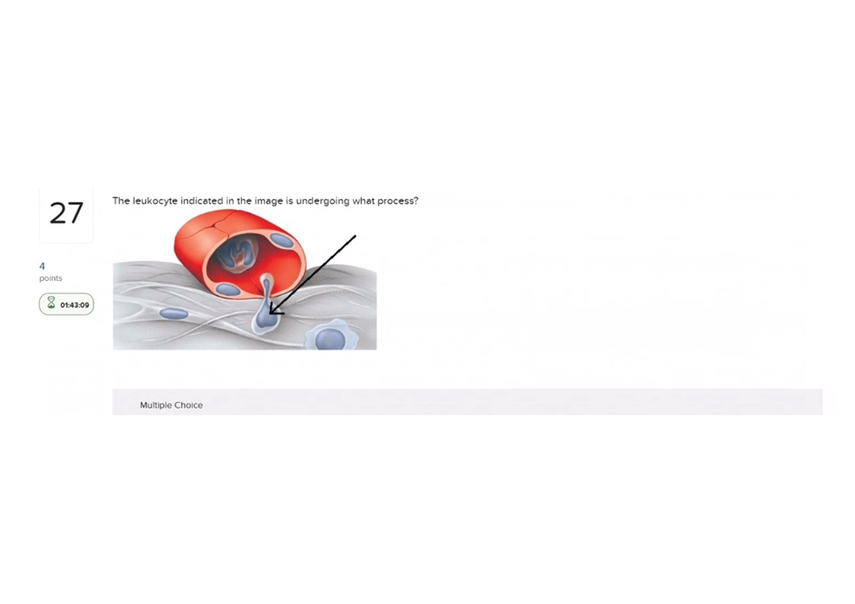
A. Diapedesis
Diapedesis (transmigration): the cell is shown squeezing through the vessel wall (leaving the bloodstream), which is diapedesis (also called extravasation).
B. Phagocytosis
Phagocytosis: phagocytosis is ingestion of particles/pathogens by a phagocyte; the image shows movement out of a vessel, not engulfment of material.
C. Chemotaxis
Chemotaxis: Incorrect (related but not the pictured action) -chemotaxis is directed movement toward chemical signals; a leukocyte may chemotax once in the tissue, but the image specifically shows the mechanical passage through the endothelium (diapedesis).
D. Margination
Margination/Rolling: margination/rolling are earlier steps along the endothelium where leukocytes slow and adhere; the image shows a cell already squeezing through the wall, which is the next step (diapedesis).
Full Explanation
A. Diapedesis (transmigration): the cell is shown squeezing through the vessel wall (leaving the bloodstream), which is diapedesis (also called extravasation).
B. Phagocytosis: phagocytosis is ingestion of particles/pathogens by a phagocyte; the image shows movement out of a vessel, not engulfment of material.
C. Chemotaxis: Incorrect (related but not the pictured action) -chemotaxis is directed movement toward chemical signals; a leukocyte may chemotax once in the tissue, but the image specifically shows the mechanical passage through the endothelium (diapedesis).
D. Margination/Rolling: margination/rolling are earlier steps along the endothelium where leukocytes slow and adhere; the image shows a cell already squeezing through the wall, which is the next step (diapedesis).